Mitosis
Mitosis is the process of dividing chromosomes during cell division in eukaryotic cells. Mitosis is followed by cytokinesis, the splitting of the cytoplasm. In cell division, a parent cell splits, producing two daughter cells that are identical to the parent. Eukaryotic unicellular organisms like the protist Amoeba use cell division in the production of new individuals, propagating their species. Multicellular eukaryotic organisms, including plants, animals, and fungi, rely on cell division to grow larger by adding new cells. They also use cell division to repair injured or worn-out tissues by replacing damaged cells with new cells.
The function of mitosis is to divide a cell’s nucleus with its chromosomes into two daughter cell nuclei, each of which inherits the same number of chromosomes as the parent cell. Consider mitosis in human cells, each of which contains forty-six chromosomes.
How does a parent cell with forty-six chromosomes divide to yield two daughter cells each with forty-six chromosomes identical to those of the parent? The eukaryotic parent cell first copies, or replicates, its chromosomes prior to mitosis. Rather than ninety-two chromosomes, however, this replication process yields forty-six chromosomes, each composed of two parts, called sister chromatids, that are genetically identical to each other. The sister chromatids are connected to each other at a point called the centromere.
During mitosis, the nuclear envelope dissolves, and sister chromatids separate at the centromere, becoming two individual daughter chromosomes, each now with only one chromatid. By the end of mitosis, these daughter chromosomes are segregated from each other to opposite poles of the cell and become enclosed within two separate daughter nuclei. Following mitosis, cytokinesis divides the cell into two, with two sets of organelles and two daughter nuclei, forming two separate but identical cells.
Specifics of Mitosis
Mitosis is a continuous process that is often divided into four sequential phases known as prophase, metaphase, anaphase, and telophase. These phases can be distinguished through microscopic analysis. Several critical steps in mitosis are controlled by phosphorylation or dephosphorylation of proteins.
Prophase. Prior to mitosis, chromosomes appear in the nucleus as a tangled mass of thin strands (chromatin) and are not distinguishable from each other as separate entities. During prophase, the chromosomes condense into shorter and thicker rod like structures that can be easily seen to consist of two sister chromatids connected by a centromere. This is thought to be driven by addition of phosphate groups to the histone proteins of the chromosome.
Prophase
Another major event in prophase is the organization of what is known as the mitotic spindle. This too is thought to be driven by phosphorylation. Prior to mitosis, a special area of the cytoplasm near the nucleus, known as the centrosome, contains a pair of small cylindrical bodies called centrioles. The centriole pairs replicate and then the two pairs of centrioles begin to move with their centrosomes to opposite poles of the cell. During prophase, they continue their migration to the cell’s poles and organize parts of the cell’s cytoskeleton (the scaffold that maintains the cell’s shape) into the mitotic spindle. The spindle consists of microtubules that reach from each centriole pair across the cell toward the other pair.
Prometaphase
On either side of the centromere that connects the two sister chromatids of each chromosome, specialized complexes of proteins known as kinetochores form. These act as attachment points between chromosomes and the spindle fibers that are part of the mitotic spindle. Through these attachments, the spindle is able to physically move the chromosomes to opposite poles of the spindle. By the end of prometaphase, the nuclear membrane surrounding the chromosomes begins to break down (triggered by phosphorylation of membrane proteins) and the spindle fibers pull the chromosomes by their kinetochore attachments so that the chromosomes align at the midpoint between the spindle poles.
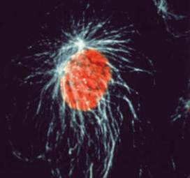
Metaphase. During metaphase the chromosomes are fully aligned end to end at the cell’s midline at what is known as the metaphase plate. Each kinetochore is attached to spindle fibers emanating from centrioles at opposite poles.

Metaphase
Anaphase. The attachments between sister chromatids to each other split during anaphase, producing single-chromatid chromosomes. This is triggered by destruction of the phosphorylating proteins discussed earlier. For each pair of single chromatid chromosomes, one of the pair is pulled toward each of the two spindle poles. Meanwhile, the distance between the spindle poles also increases.
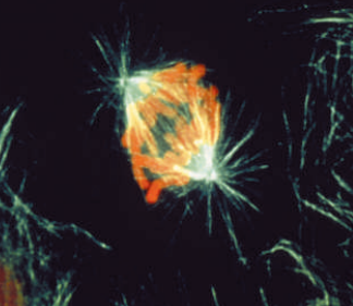
Anaphase
Telophase. During telophase, the nuclear membranes are dephospho- rylated and begin to reform around the two sets of chromosomes at either pole, enclosing and separating them from the rest of the cytoplasm. The mitotic spindle disappears. The chromosomes decondense and become thinner and more difficult to distinguish from each other. Cytokinesis begins the process of separating the two daughter cells and is nearly complete by the end of telophase. The end result is the production of two new cells that are genetically identical to each other and to the parent cell.
Differences between Plants and Animals
Plants use a similar process with a few differences. For example, although a plant cell creates a mitotic spindle and has a centrosome, it lacks centrioles. The other major difference in plants is the way in which cytokinesis occurs. In animal cells, the plasma membrane pinches in along the midline of the cell, creating a cleavage furrow that will separate the cytoplasm in two. Plant cells have rigid cell walls that prevent this. Instead, they use two different approaches for cytokinesis. The plasma membrane and cell wall grow inward together, eventually separating the parent cell into two. Alternatively, the cell wall that will separate the two daughter cells starts growing in the middle of the cell between the two nuclei and continues toward the periphery. This is known as the cell plate. It continues growing until its edges reach the cell’s outer surface, separating the parent cell into two daughter cells.
References
Mader, Sylvia S. Biology, 6th ed. Boston: WCB McGraw-Hill, 1998.
McFadden, Carol H., and William T. Keeton. Biology: An Exploration of Life. New York: W. W. Norton and Company, Inc., 1995.