آخر المواضيع المضافة

 النبات
الحيوان
الأحياء المجهرية
علم الأمراض
التقانة الإحيائية
التقنية الحيوية المكروبية
التقنية الحياتية النانوية
علم الأجنة
الأحياء الجزيئي
علم وظائف الأعضاء
الغدد
المضادات الحيوية
النبات
الحيوان
الأحياء المجهرية
علم الأمراض
التقانة الإحيائية
التقنية الحيوية المكروبية
التقنية الحياتية النانوية
علم الأجنة
الأحياء الجزيئي
علم وظائف الأعضاء
الغدد
المضادات الحيوية| Cell Wall of Plants |


|
|
Read More
Date: 6-11-2016
Date: 20-10-2016
Date: 17-10-2016
|
Cell Wall of Plants
Almost all plant cells have a cell wall; only sperm cells of some seed plants lack one. It is often regarded as simply an inert secretion, providing only strength and protection to the protoplasm inside. However, considerable metabolism occurs in the wall, and it should therefore be considered a dynamic, active organelle.
The cell wall contains a considerable amount of the polysaccharide cellulose (see fig 1a). Adjacent, parallel cellulose molecules crystallize into an extremely strong microfibril 10 to 25 nm wide. Numerous microfibrils are wound around the cell, completely covering the plasma membrane. Each cellulose polymer grows only at one end, where a complex of enzymes adds new glucose residues, one molecule at a time (Fig. 1b). The enzymes are intrinsic proteins of the plasma membrane, and as they add new sugars to the chain, the enzymes float forward in the membrane (the chain is too heavy to be pushed backward). New cellulose molecules can be added only on the inner side of the wall, adjacent to the plasma membrane.
Cellulose microfibrils are bound together by other polysaccharides called hemicelluloses, which are produced in dictyosomes and brought to the wall by dictyosome vesicles. Hemicelluloses are deposited between the cellulose microfibrils and bind chemically to the cellulose, producing a solid structure that resembles reinforced concrete (Fig. 2). In multicellular plants, the wall of one cell is glued to the walls of adjacent cells by an adhesive layer, the middle lamella, composed of a third class of polysaccharides, pectic substances.
All plant cells have a thin wall called the primary cell wall. In certain cells that must be unusually strong, the protoplast deposits a secondary cell wall between the primary wall and the plasma membrane. The secondary wall is usually much thicker than the primary wall and is almost always impregnated with the compound lignin, which makes the wall even stronger than hemicelluloses alone can make it. Lignin resists chemical, fungal, and bacterial attack. Both primary and secondary cell walls are permanent; once deposited, they are almost never degraded or depolymerized, as can be done with microtubules and microfilaments.
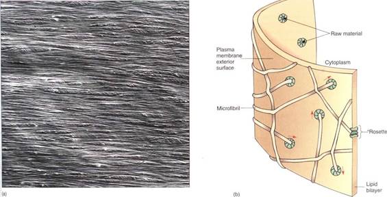
FIGURE 1: (a) Two layers of cellulose microfibrils are visible; other layers are present deeper in the wall. Each layer provides strength in the direction parallel to the microfibrils. Other wall components have been dissolved away to reveal the cellulose (X 5000). (Courtesy of Dr. Shun Mizuta, Kochi University, Japan) (b) Cellulose-synthesizing enzymes are large proteins embedded in the plasma membrane; several proteins form a cluster called a rosette. Sugar monomers are absorbed by the enzymes of the membrane inner face, then passed across the membrane and polymerized into cellulose on the outer face. The growing microfibril extends out from the rosette and lies against microfibrils already in place. Because the microfibril cannot push into the wall, the rosette is pushed through the membrane in the direction of the arrows as the microfibril grows.
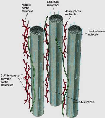
FIGURE 2: Although cellulose microfibrils lie close together, they do not bond with each other, so a wall of pure cellulose is weak. Hemicelluloses and pectins are short, branched molecules that interact with several adjacent microfibrils, linking them together and forming a solid threedimensional mesh. The relative amounts of these components can vary, resulting in some walls that are flexible, others that are rigid; some strong, others weak.


|
|
|
|
دراسة يابانية لتقليل مخاطر أمراض المواليد منخفضي الوزن
|
|
|
|
|
|
|
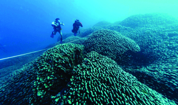
اكتشاف أكبر مرجان في العالم قبالة سواحل جزر سليمان
|
|
|
|
|
|
|
اتحاد كليات الطب الملكية البريطانية يشيد بالمستوى العلمي لطلبة جامعة العميد وبيئتها التعليمية
|
|
|