آخر المواضيع المضافة

 النبات
الحيوان
الأحياء المجهرية
علم الأمراض
التقانة الإحيائية
التقنية الحيوية المكروبية
التقنية الحياتية النانوية
علم الأجنة
الأحياء الجزيئي
علم وظائف الأعضاء
الغدد
المضادات الحيوية
النبات
الحيوان
الأحياء المجهرية
علم الأمراض
التقانة الإحيائية
التقنية الحيوية المكروبية
التقنية الحياتية النانوية
علم الأجنة
الأحياء الجزيئي
علم وظائف الأعضاء
الغدد
المضادات الحيوية| Cardiovascular System |


|
|
Read More
Date: 18-7-2021
Date: 1-8-2021
Date: 18-7-2021
|
Cardiovascular System
The cardiovascular system consists of the heart and blood vessels that circulate blood throughout the body (Fig. 1).
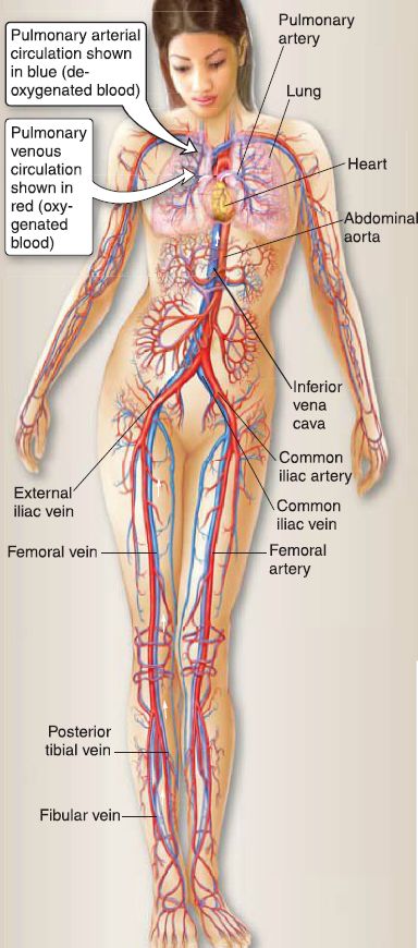
Figure 1: Cardiovascular system.
A. Heart
The heart consists of two muscular pumps that divide the blood circulation into two circuits (Fig. 2). In the pulmonary circulation, the right ventricle pumps low-oxygen blood into the lungs via the pulmonary arteries where the blood is oxygenated and then returned to the left atrium of the heart via the pulmonary veins. In the systemic circulation, the left ventricle pumps highly oxygenated blood through the systemic arteries to distribute oxygen and nutrients throughout the body. Low-oxygen blood is returned to the right atrium of the heart via systemic veins.
B. Blood vessels
The three general types of blood vessels are arteries, capillaries, and veins, which distribute blood throughout the body. For a summary of blood vessel types, see Figure 5.
Systemic blood flow follows a particular pathway: Elastic arteries ➔ Muscular arteries ➔ Arterioles ➔ Metarterioles ➔ Capillary bed ➔ Postcapillary venules ➔ Muscular venules ➔ Collecting venules ➔ Veins of increasing diameter (named veins)

Figure 2: Pulmonary and systemic circulations. Red is oxygenated blood; blue is deoxygenated blood.
1. Arteries: The vascular wall of arteries consists of three concentric layers ("tunics"): tunica intima, tunica media, and tunica adventitia (Fig. 3). The tunica intima is the innermost layer and consists of the endothelium, basal lamina, subendothelial loose connective tissue, and an internal elastic lamina. The tunica media is the middle layer and consists of smooth muscle cells, type Ill collagen fibers, elastic fibers, and an external elastic lamina. The tunica adventitia is the outermost layer and consists of fibroblasts, type I collagen fibers, and scattered elastic fibers. The tunica adventitia of large arteries contains small blood vessels (vasa vasorum), small postganglionic sympathetic nerve bundles (nervi vascularis), and small lymph vessels. In the circulatory system, elastic arteries gradually transition to large muscular arteries (i.e.,no line demarcates them). In the region of transition, the amount of elastic fibers in the tunica media decreases, whereas the amount of smooth muscle whereas the amount of smooth muscle in the tunica media increases.
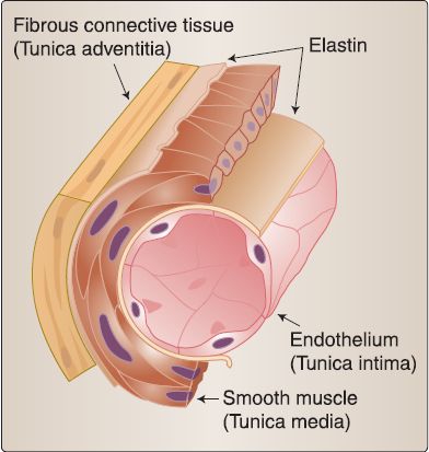
Figure 3: Blood vessel structure.
a. Elastic artery: Elastic arteries function primarily as conduction arteries; that is, they conduct blood from the heart to the muscular arteries. Elastic arteries are distinguished by a tunica media with a prominent elastic fiber component that consists of concentric layers of fenestrated elastic lamellae (or sheets).
They receive blood under high systolic pressure from the heart and keep the blood circulating while the heart pumps intermittently. Elastic arteries distend during systole and recoil during diastole. Examples include the pulmonary trunk, aorta, common carotid arteries, subclavian arteries, and common iliac arteries.
b. Muscular artery: Muscular arteries function primarily as distribution arteries. They have a tunica media with a prominent smooth muscle component that controls the distribution of blood to organs and tissues of the body. Examples include the axillary, ulnar, radial, and femoral arteries.
c. Arteriole: Arterioles function primarily as resistance vessels. They regulate blood flow to the capillary beds. The contraction of the one or two layers of smooth muscle cells in the tunica media increases the vascular resistance and thereby reduces blood flow to the capillary bed. The arterioles offer the greatest resistance to the flow of blood from the heart to the peripheral tissues and therefore play a role in the regulation of arterial blood pressure.
d. Metarteriole: A metarteriole is the terminal branch of the arterial system and flows directly into the capillary bed. It has a thickened smooth muscle cell layer that acts as a precapillary sphincter, regulating blood flow to the capillary bed.
2. Capillaries: The vascular wall of a capillary consists of an endothelium, a basal lamina, and pericytes. A pericyte is a cell that has contractile properties and can proliferate in response to tissue injury to act as a stem cell during angiogenesis. The capillary forms a small tube with a diameter that allows for the passage of red blood cells one at a time. Capillaries function primarily as exchange vessels. The capillary is the principle site of exchange of water, oxygen, carbon dioxide, glucose, amino acids, proteins, metabolites, and waste products between the blood and cells.
a. Continuous capillary: A continuous capillary consists of a single layer of endothelial cells joined by a zonula occludens (tight junction). It is surrounded by a continuous basal lamina and is found in the lung, muscle, thymus (blood-thymus barrier), nervous system (blood-brain barrier), connective tissue, and exocrine glands.
b. Fenestrated capillary: As shown in Figure 4, a fenestrated capillary consists of a single layer of endothelial cells joined by a fascia occludens (a tight junction that extends only partially around the perimeter of the cell creating small slit-like intercellular spaces). It is surrounded by a continuous basal lamina and has numerous fenestrae (or pores). The fenestrae are generally bridged by a diaphragm. They are found in the kidney (except that in the kidney glomerulus the fenestrated capillary has no diaphragms), lamina propria of the intestine, choroid plexus of the brain, choriocapillaris of the eye, and endocrine glands.
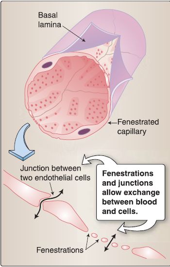
Figure 4: Capillary wall structure.
c. Discontinuous capillary (sinusoid): A discontinuous capillary consists of a single layer of endothelial cells joined by a fascia occludens. Like fenestrated capillaries, it has numerous fenestrae, but its basal lamina is discontinuous. They are found in the liver, bone marrow, and spleen.
3. Veins: Like arteries, the vascular wall of veins consists of three concentric tunics. The tunica media of veins is thinner than that of arteries, and the tunica adventitia is thicker. Veins have valves that are projections of the tunica intima. The transport of blood back to the heart via veins depends on the contraction of skeletal muscles and valves that ensure one-way flow of blood.
a. Postcapillary venule: The vascular wall of a postcapillary venule (-0.2 mm in diameter) consists of an endothelium, a basal lamina, and pericytes. The postcapillary venule is the principal site of action for vasoactive agents (e.g., histamine, serotonin) resulting in the extravasation of fluid during inflammation and allergic reactions, migration of inflammatory leukocytes (i.e., diapedesis), and migration of lymphocytes to repopulate the lymph node.
b. Muscular and collecting venules: The vascular wall of a venule consists of three tunics.
c. Medium- and large-sized veins: Medium- and large-sized veins are high-capacitance or reservoir vessels that contain about 70% of the total blood volume.
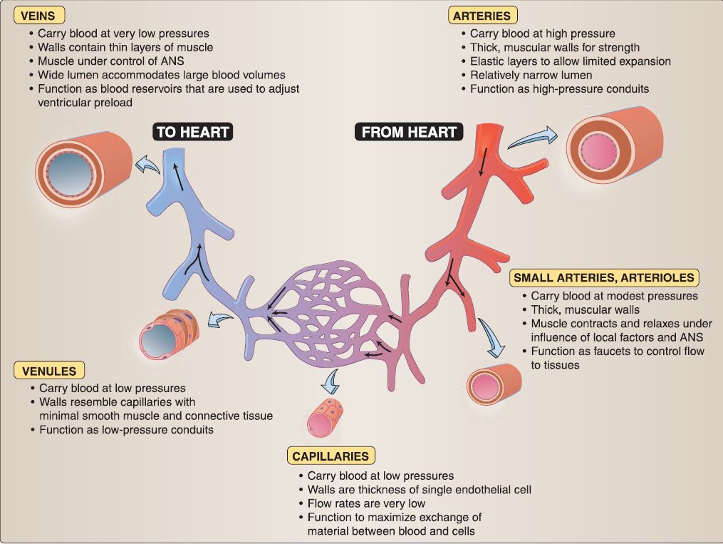 Figure 5: Organization of arteries and veins.


|
|
|
|
دراسة يابانية لتقليل مخاطر أمراض المواليد منخفضي الوزن
|
|
|
|
|
|
|
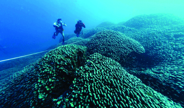
اكتشاف أكبر مرجان في العالم قبالة سواحل جزر سليمان
|
|
|
|
|
|
|
اتحاد كليات الطب الملكية البريطانية يشيد بالمستوى العلمي لطلبة جامعة العميد وبيئتها التعليمية
|
|
|