آخر المواضيع المضافة

 النبات
الحيوان
الأحياء المجهرية
علم الأمراض
التقانة الإحيائية
التقنية الحيوية المكروبية
التقنية الحياتية النانوية
علم الأجنة
الأحياء الجزيئي
علم وظائف الأعضاء
الغدد
المضادات الحيوية
النبات
الحيوان
الأحياء المجهرية
علم الأمراض
التقانة الإحيائية
التقنية الحيوية المكروبية
التقنية الحياتية النانوية
علم الأجنة
الأحياء الجزيئي
علم وظائف الأعضاء
الغدد
المضادات الحيوية| Pelvic Vasculature |


|
|
Read More
Date: 21-7-2021
Date: 12-7-2021
Date: 5-11-2015
|
Pelvic Vasculature
At the aortic bifurcation, right and left common iliac arteries course laterally before terminating as internal and external iliac arteries bilaterally (Figs. 1 and 2). The external iliac arteries continue toward the anterior thigh. The internal iliac arteries course medially, divide into posterior and anterior divisions, and serve as the main blood supply to pelvic and perinea! structures. Venous drainage of pelvic and perinea! structures occurs through tributaries of the iliac system, which then drain into the caval circulation. External and internal iliac veins unite to form common iliac veins, which merge to form the inferior vena cava at approximately the L4-L5 level.
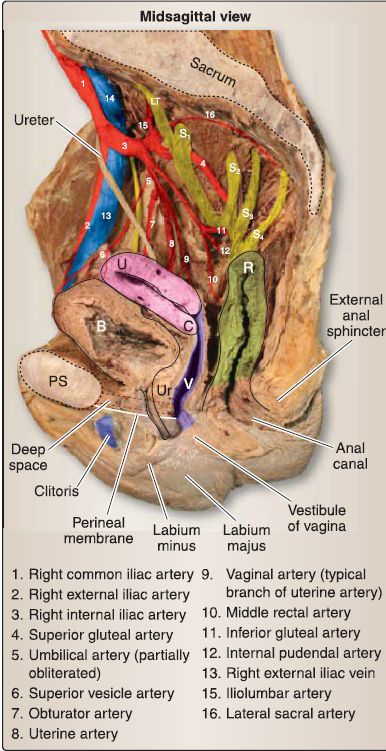
Figure 1: Female pelvic vasculature and of vagina sacral plexus. Parietal peritoneum
removed. B = bladder, C = cervix, LT = lumbosacral trunk, PS = pubic symphysis, R = rectum, U = uterus, Ur = urethra, V = vagina.
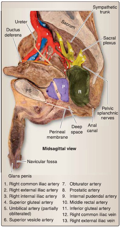
Figure 2: Male pelvic vasculature and sacral plexus. Parietal peritoneum removed. B = bladder, P = prostate gland, PS = pubic symphysis, R = rectum, Ur= urethra.
A. Internal iliac posterior division
Three branches arise from the posterior division of the internal iliac artery. These branches primarily supply gluteal and pelvic wall structures.
1. lliolumbar: This artery provides muscular branches to iliacus, psoas major, and quadratus lumborum muscles.
2. Lateral sacral: This branch can present as paired arteries and courses interiorly along the sacrum to enter an anterior sacral foramen.
3. Superior gluteal: The largest of the three posterior division branches, this artery typically passes between the lumbosacral trunk (L4-L5) and S1 to exit the pelvis through the greater sciatic foramen. It enters the gluteal region superior to the piriformis and supplies gluteus maximus, minimus, and medius and tensor fasciae latae.
B. Internal iliac anterior division
Multiple branches arise from the anterior division, which supply thigh, perinea!, gluteal, and pelvic structures.
1. Obturator: This artery exits the pelvis through a small opening in the obturator membrane to enter the medial thigh and gives off a small acetabular branch to the head of the femur.
2. Umbilical: The distal end of this artery becomes obliterated after birth to form the medial umbilical ligament, and its proximal end gives off the artery to ductus deferens and multiple superior vesical branches to the bladder.
3. Uterine: This artery passes along the side of uterus to the broad ligament base, where it is crossed inferiorly by the ureter and supplies the uterus, fallopian tubes, and upper vagina.
a. Vaginal: Commonly a branch of the uterine artery, this artery supplies the vaginal wall.
4. Inferior vesical: A homologue to the vaginal artery in females, this artery supplies the bladder, seminal glands, and prostate.
5. Middle rectal: This artery may share a common stem with the internal pudenda! or inferior vesicle and contributes to collateral blood flow to the rectum.
6. Internal pudenda!: This artery often shares a common stem with the inferior gluteal artery. It exits the pelvis through the greater sciatic foramen, then enters the ischioanal fossa through the lesser sciatic foramen. It gives off inferior gluteal branches before supplying structures of the perineum.
7. Inferior gluteal: This artery exits the pelvis through the greater sciatic foramen and enters the gluteal region inferior to piriformis. It supplies the gluteus maximus primarily.
C. Venous drainage
A series of pelvis venous plexuses surround pelvic viscera within the true (lesser) pelvis. These plexuses primarily drain into tributaries of internal iliac veins, although portacaval anastomoses are present in the rectum. Venous drainage from the gonads (testes and ovaries) terminates in the caval system by way of gonadal veins (testicular and ovarian). Right and left gonadal veins drain into the inferior vena cava and left renal vein, respectively .


|
|
|
|
للعاملين في الليل.. حيلة صحية تجنبكم خطر هذا النوع من العمل
|
|
|
|
|
|
|
"ناسا" تحتفي برائد الفضاء السوفياتي يوري غاغارين
|
|
|
|
|
|
|
ملاكات العتبة العباسية المقدسة تُنهي أعمال غسل حرم مرقد أبي الفضل العباس (عليه السلام) وفرشه
|
|
|