

 النبات
 الحيوان
 الأحياء المجهرية
 علم الأمراض
 التقانة الإحيائية
 التقنية الحيوية المكروبية
 التقنية الحياتية النانوية
 علم الأجنة
 الأحياء الجزيئي
 علم وظائف الأعضاء
 الغدد
الغدد
 المضادات الحيوية
|
Read More
Date: 16-2-2016
Date: 16-2-2016
Date: 15-2-2016
|
Enzyme-Linked Immunosorbent Assay (ELISA(
Enzyme-linked immunosorbent assay (ELISA) is a technique for quantifying a substance by use of an antibody specific for that substance that has been conjugated to an enzyme. The original ELISA was developed along the lines of a solid-phase radioimmunoassay, substituting an enzyme for a radioactive label (1, 2). Regardless of the experimental design used for the immunological reactions, the end result is that the antibody–enzyme conjugate is immobilized. Enzymatic activity is assayed in a subsequent reaction, and the activity is related to the quantity of analyte (antigen or antibody( present in a test sample by comparison to a set of controls of known concentration.
A key reagent for all ELISAs is the enzyme conjugate. In most cases, the enzyme is crosslinked covalently to an antibody, but experimental designs with enzyme-labeled antigen are also used. Indeed, the ELISA method is adaptable to any ligand and receptor pair. The most common enzymes used are alkaline phosphatase and horseradish peroxidase. These proteins are stable to the crosslinking procedures, and they catalyze reactions with highly chromogenic substrates, leading to very sensitive detection limits. Optical absorbance is the most common method of measuring enzyme activity, but other detection technologies are also used, such as fluorescence or chemiluminescence. Covalent linkage to a chromatography matrix was used to immobilize the first “immunosorbent,” but use of plastic multiwell microtiter plates is now nearly universal, as these allow simultaneous monitoring of scores or hundreds of enzymic reactions in automated instruments.
The number of experimental formats used for ELISA is immense. This discussion is limited to the simplest and most useful assay designs.
1. Competitive ELISA
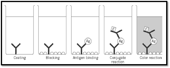
The first reported ELISA was a competition assay, in which adding an unlabeled competitor molecule reduced the amount of enzyme conjugate that could be immobilized (1). This remains an extremely common approach. The method begins with adsorption of antigen to the surface of a microtiter plate (Fig. 1). This non-specific adsorption occurs spontaneously, simply by allowing the antigen solution to stand in the wells of the plate. The linkage to the plate is noncovalent, but essentially irreversible, and the adsorbed antigen is seldom released during subsequent reactions and washing steps. Often a blocking step follows, in which a concentrated solution of a protein such as serum albumin is used to coat the remaining plastic surface, thereby preventing later adsorption of assay components. The plate is washed with a neutral buffer. The antibody solution of unknown concentration, and a series of controls, consisting of known quantities of the same antibody, are mixed with a fixed amount of the antibody–enzyme conjugate and added to different wells of the plate. The conjugate binds to the immobilized antigen, but so does the unconjugated antibody, and the binding of each is mutually exclusive. Thus, the higher the concentration of unconjugated antibody, the less the amount of enzyme immobilized. After allowing an interval for the antigen to be bound, the plate is again washed, and the addition of substrate initiates the enzymatic reaction. The rate of product release, or the accumulation of product after a fixed time, is determined for the unknown analyte and controls. Reactions to which no unlabeled competitor was added will have the highest absorbance, and are taken as a 100% response. Controls lacking conjugate, or lacking antigen, or to which a saturating amount of competitor was added, give the lowest absorbance. This value, reflecting background enzyme binding and the uncatalyzed chromogenic reaction, is taken as a 0% response. Data from the other controls are plotted to give a calibration curve, using a logarithmic concentration scale on the abscissa and an absorbance or percentage scale on the ordinate (Fig. 2a). The response from the unknown is compared to the calibration curve, and the corresponding concentration is read off the abscissa.

Figure 1. Competitive ELISA.

Figure 2. ELISA standard curves.
A shortcoming of this competitive design is that the response from small amounts of analyte may be almost as high as the 100% control. The change in reading due to the analyte is thus read as the difference between two large numbers. To be considered precise, the minimum readable difference must be much larger than the standard deviation of the highest point on the calibration curve, a fact that inherently limits the sensitivity of competitive ELISAs. The two noncompetitive designs that follow circumvent this limitation. The response in these assays is proportional to the analyte added; hence even a low reading may be detectable against the background level of enzymatic activity (Fig. 2b).
2. Indirect ELISA
The enzyme conjugate in an indirect ELISA (3) is an anti-antibody; the antibody that actually recognizes that the antigen is unlabeled. Quantification of the unlabeled antibody is normally the goal sought, and the indirect ELISA is most commonly used to assess the seropositivity of subjects who have potentially been exposed to pathogens. In an indirect ELISA, antigen is immobilized and the plate blocked as above (Fig. 3). The test sample is added, along with controls consisting of antibodies of the same specificity, species, and isotype as the test sample. After an interval allowed for antigen binding, the plate is washed and the conjugate added. The antibody used to make the conjugate reagent is typically a polyclonal preparation specific for constant regions of the antibody in the test sample. Finally, enzymic activity is assayed as above. Controls are plotted on a linear or log–log scale (Fig. 2b). In this case, the response increases with increasing amount of analyte and does not necessarily reach a plateau. Reaching a plateau at high analyte concentration may indicate that the binding capacity of the unlabeled antibody in the sample exceeds the quantity of conjugate added, or more trivially, that the optical density after the color reaction is simply too great to be measured. The concentration of analyte in the test sample is interpolated from the calibration curve, as above.

Figure 3. Indirect ELISA.
3. Sandwich ELISA for Antigen
This ELISA method (4) is conceptually similar to the indirect ELISA described above, but the analyte is usually a nonimmunoglobulin antigen that becomes “sandwiched” between an unlabeled immobilizing antibody and an antibody–enzyme conjugate (Fig. 4). The procedure begins with adsorption of a specific antibody to the surface of the assay plate. The plate is washed, and the test sample is added, along with controls consisting of known quantities of the same antigen. The plate is again washed, and the antibody–enzyme conjugate is added. This reagent binds to the immobilized antigen and hence becomes immobilized itself. A crucial aspect of the design of a sandwich assay is selection of an immobilizing antibody and a conjugate antibody that together will actually allow a “sandwich” to form. Obviously, two monoclonal antibodies that recognize the same epitope will exclude each other's binding. Use of a monoclonal immobilizing antibody is advantageous, however, since it occludes only a single epitope, leaving the remaining surface of the antigen available for deposition of the antibody–enzyme conjugate. After the conjugate is allowed to bind, the tray is washed a final time, and substrate is added to initiate the color reaction. A rising calibration curve again results, from which sample concentration is interpolated as above.

Figure 4. Sandwich ELISA.
4. Affinity Determination
Many authors have equated the analyte concentration at the 50% response point in the competitive ELISA with the equilibrium dissociation constant for an antibody–antigen interaction. This approximation is invalid. However, ELISA can be used to measure the equilibrium constant if the indirect ELISA method is applied after equilibrium is reached (5). The antibody and antigen in question are mixed in a series of different ratios, and the solutions are allowed to come to equilibrium. The equilibrium mixtures are then transferred to an ELISA plate that has been coated with the antigen. Standards with known amounts of free antibody are run in parallel. Free antibody will bind to the immobilized antigen. It is essential that only a small fraction of the total available amount of free antibody become bound to the plate, so that the assay does not lead to an apparent shifting of the equilibrium point (6). The plate is washed, and an antibody–enzyme conjugate that binds to the immobilized antibody is added. The plate is washed again, substrate solution added, and the amount of enzyme activity determined. The color response from the reactions with the equilibrium mixtures is compared to that from the controls, and the relative response is equated to the concentration of free antibody at equilibrium. A correction can be applied to allow for the multivalency of the antibody (7). Using the measurements of free antibody and knowledge of the total antibody and antigen present, one can infer the concentrations of free and bound antigen and antibody. These data can then be graphed on a Scatchard Plot, or numerical analysis can be used to determine the association equilibrium constant for the antibody–antigen interaction.
References
1. E. Engvall and P. Perlman (1971) Immunochemistry 8, 871–874.
2. B. K. van Weemen and A. H. W. M. Schnuurs (1971) FEBS Lett. 15, 232–237.
3. E. Engvall and P. Perlmann (1972) J. Immunol. 109, 129–135.
4. L. Belanger, C. Sylvestre, and D. Dufour (1973) Clin. Chim. Acta 48, 15–18.
5. B. Friguet, A. F. Chaffotte, L. Djavadi-Ohaniance, and M. E. Goldberg (1985) J. Immunol. Meth. 77, 305-319.
6. S. Hetherington (1990) J. Immunol. Meth. 131, 195–202.
7. F. S. Stevens (1987) Mol. Immunol. 24, 1055–1060.


|
|
|
|
تفوقت في الاختبار على الجميع.. فاكهة "خارقة" في عالم التغذية
|
|
|
|
|
|
|
أمين عام أوبك: النفط الخام والغاز الطبيعي "هبة من الله"
|
|
|
|
|
|
|
قسم شؤون المعارف ينظم دورة عن آليات عمل الفهارس الفنية للموسوعات والكتب لملاكاته
|
|
|