آخر المواضيع المضافة

 النبات
الحيوان
الأحياء المجهرية
علم الأمراض
التقانة الإحيائية
التقنية الحيوية المكروبية
التقنية الحياتية النانوية
علم الأجنة
الأحياء الجزيئي
علم وظائف الأعضاء
الغدد
المضادات الحيوية
النبات
الحيوان
الأحياء المجهرية
علم الأمراض
التقانة الإحيائية
التقنية الحيوية المكروبية
التقنية الحياتية النانوية
علم الأجنة
الأحياء الجزيئي
علم وظائف الأعضاء
الغدد
المضادات الحيوية| Cryoelectron Microscopy |


|
|
Read More
Date: 9-5-2016
Date: 25-12-2015
Date: 25-5-2021
|
Cryoelectron Microscopy
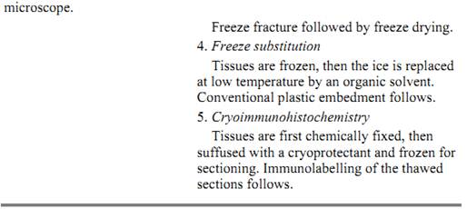
Cryoelectron microscopy can be divided into two classes, depending on whether the specimen is hydrated or dry. Table 1 provides a brief description of the possible methods of specimen preparation. In each method, the specimen is initially frozen and can subsequently be observed in a cryoelectron microscope, or can be further preserved by drying or making a replica for observation in a conventional electron microscope.
Table 1. Methods of Specimen Preparation in Cryoelectron Microscopy

The development of techniques for visualizing frozen hydrated tissues and macromolecules has been a boon for studying structures in a state that closely approximates their native condition (1). Because freezing takes place very rapidly, these techniques offer the ability to examine dynamic events with high time resolution. For example, the structure of the open-channel form of the acetylcholine receptor was elucidated from membrane crystals that were briefly activated (< 5 ms) with droplets containing acetylcholine (2). Small conformational changes triggered by acetylcholine were detected in the frozen hydrated crystals. The spatial resolution of periodic structures, eg, protein crystals, can also be high, extending to 3.3 Å (3; Perkins, unpublished results). Moreover, these structures can be examined unstained by utilizing the intrinsic contrast difference between the biological specimen and vitreous ice. Thus, the effects of solvent composition on the structure and aspects of interacting components can be examined (2).
Fundamental to examining frozen hydrated specimens is the preservation of the environment by rapid freezing (4). Rapid freezing causes the water of the specimen and its environment to be trapped in a vitreous (amorphous or glass-like) state. This vitreous state prevents damage to the specimen caused by the formation of ice crystals when the specimen is frozen more slowly. The specimens preserved in thin films of vitreous ice generally retain a high degree of structural integrity. However, it is crucial to maintain the specimen at below ~ –140° C because vitreous ice tends to crystallize above this temperature (4.(
A wide range of frozen hydrated specimens has been examined in cryoelectron microscopes. Most studies have concentrated on macromolecules and their assemblies hydrated and frozen in thin films. To a lesser extent cryosections of biological tissues have also been studied (5-9), but the difficulty of freezing of bulk material without formation of ice crystals has hampered progress. The first published results obtained of macromolecules were from viruses, fairly robust particles (10). Early work produced 3-D reconstructions at relatively low resolution, ~ 2–3 nm (10-13), although certain viruses produced data to ~ 1 nm resolution (14). Important new information about viral structures was obtained even at lower resolution. Recent work has extended the resolution of complex viruses beyond 1 nm (15, 16). By combining cryoelectron microscopy with X-ray crystallography, near atomic resolution detail of the overall architecture of large complexes and of the interactions between the components can be obtained. An instructive example is the 3-D structure of rhinovirus 14 complexed with Fab fragments determined at 0.4 nm resolution (17).
Although the contrast in frozen hydrated specimens is low compared to negatively stained specimens, the signal can be significantly boosted by averaging many identical particles together using Fourier averaging for crystals (2, 17) and correlational averaging for single particles as discussed below (15, 16, 18). Vitreous ice embedment is particularly useful for visualizing the specimen interior compared to negative stain, which accumulates around areas accessible to the aqueous phase, thus usually showing only the molecular envelope. Hence, in general only those portions of integral membrane proteins extending beyond the lipid bilayer are observed in negative stain. However, exceptions to this limitation of negatively stained membrane proteins have been found (19-22). In certain instances, frozen hydrated crystals of membrane proteins do not provide resolution as high as the same crystals in negative stain (23). Vitreous ice embedment of delicate specimens, such as chromosomes or DNA fragments (9, 24, 25) has provided impressive results, since this type of specimen is commonly poorly preserved in other embedment media. Another example is the direct visualization by cryoelectron microscopy of A-, P-, and E-site transfer RNAs in the E. coli ribosome (26).
Cryofixation of tissues is an alternative to conventional chemical fixation of subcellular ultrastructure. Vitrification of ice can frequently be achieved to a depth of 10–20 µm when tissues are frozen by slamming them against a polished copper block cooled to the temperature of liquid nitrogen or liquid helium (27). High-pressure freezing can freeze samples in vitreous ice to depths as great as 100 µm, since the increase in volume going from water to ice is impaired by a high-pressure environment inhibiting the growth of ice crystals. Cryofixation is usually the choice for immunocytochemistry because it retains the antigenic properties of the specimen better than chemical fixation. With good vitrification, freeze-etching or freeze-substitution (Table 1) can provide information about structures stabilized with a mechanism different from chemical fixation.
References
1. J. Jaffe and R. M. Glaeser (1984) Ultramicroscopy 13, 373–378.
2. P. N. T. Unwin (1995) Nature 373, 37–43.
3. J. Brink, W. Chiu, and M. Dougherty (1992) Ultramicroscopy 46, 229–240.
4. J. Dubochet et al. (1982) J. Microscopy 128, 219–237.
5. J.-J. Chang et al. (1983) J. Microscopy 132, 109–123.
6. J. Dubochet et al. (1983) J. Bacteriol. 155, 381–390.
7. A. W. McDowall et al. (1983) J. Microscopy 131, 1–9.
8. A. W. McDowall et al. (1984) J. Mol. Biol. 178, 105–11.
9. A. W. McDowall, J. M. Smith, and J. Dubochet (1986) EMBO J. 5, 1395–1402.
10. J. Adrian et al. (1984) Nature 308, 32–36.
11. J. Lepault and K. Leonard (1985) J. Mol. Biol. 182, 431–441.
12. F. P. Booy et al. (1985) J. Mol. Biol. 184, 667–676.
13. R. H. Vogel et al. (1986) Nature 320, 533–535.
14. J. Lepault (1985) J. Microscopy 140, 73–80.
15. B. Bottcher, S. A. Wynne, and R. A. Crowther (1997) Nature 386, 88–91.
16. J. F. Conway et al. (1997) Nature 386, 91–94.
17. T. J. Smith (1996) Nature 383, 350–354.
18. M. van Heel, G. Harauz, and E. V. Orlova (1992) J. Struct. Biol. 116, 17–24.
19. B. Karlsson et al. (1983) J. Mol. Biol. 165, 287–302.
20. B. Bottcher, P. Graber, and E. J. Boekema (1992) Biochem. Biophys. Acta 1100, 125–136.
21. S. Karrasch et al. (1996) J. Mol. Biol. 262, 336–348.
22. G. A. Perkins et al. (1997) Biophys. J. 72, 533–544.
23. J. M. Valpuesta, R. Henderson, and T. G. Frey (1990) J. Mol. Biol. 214, 237–251.
24. J. Dubochet et al. (1994) Nature Struct. Biol. 1, 361–363.
25. J. Bednar et al. (1995) J. Mol. Biol. 254, 579–594.
26. R. K. Agrawal et al. (1996) Science 271, 1000–1002.
27. J. Dubochet (1995) Trends Cell Biol. 5, 366–369.


|
|
|
|
دراسة يابانية لتقليل مخاطر أمراض المواليد منخفضي الوزن
|
|
|
|
|
|
|
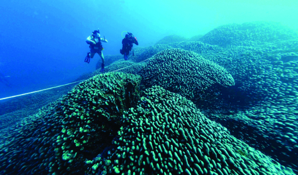
اكتشاف أكبر مرجان في العالم قبالة سواحل جزر سليمان
|
|
|
|
|
|
|
اتحاد كليات الطب الملكية البريطانية يشيد بالمستوى العلمي لطلبة جامعة العميد وبيئتها التعليمية
|
|
|