آخر المواضيع المضافة

 النبات
الحيوان
الأحياء المجهرية
علم الأمراض
التقانة الإحيائية
التقنية الحيوية المكروبية
التقنية الحياتية النانوية
علم الأجنة
الأحياء الجزيئي
علم وظائف الأعضاء
الغدد
المضادات الحيوية
النبات
الحيوان
الأحياء المجهرية
علم الأمراض
التقانة الإحيائية
التقنية الحيوية المكروبية
التقنية الحياتية النانوية
علم الأجنة
الأحياء الجزيئي
علم وظائف الأعضاء
الغدد
المضادات الحيوية| History of Human anatomy |


|
|
Read More
Date: 12-7-2021
Date: 9-7-2021
Date: 27-7-2021
|
History of Human anatomy
The term ‘anatomy’ is Greek in origin. It takes its root from ‘ana’ and ‘tome’ (ana-up; tome-cutting). Thus anatomy is the science of physical structure of an animal or plant studied by dissection. The Human Anatomy provided the necessary knowledge for surgery and medicine.
The study of human anatomy dates back to 2500 BC, when the Egyptians prepared mummies. They removed internal organs of cadavers being mummified. They also did surgery for wounds and broken bones. In India during 500 - 491 BC Susruta performed cataract operation. In 1st century AD, Celsus, a Roman physician wrote about surgical procedures.
The year 1543 AD was significant due to publication of an accurate book on Anatomy by Andreas Vesalius. In 1628 William Harvey described the functioning of heart and the movement of blood in animals. These earlier works were followed by the discovery and accurate account of each and every organ system and organs in human body. In the recent times, attempts are being made to understand the molecular architecture in every cell of our body.
The Integumentary System
The word integument means covering. The integumentary system covers the outside of the body. It protects internal structures, prevents the entry of infectious agents, reduces water loss, regulates body temperature, produces vitamin D and detects stimuli such as touch, pain and temperature. Since the integument performs several functions, it is commonly referred to as Jack of all trades.
The skin or integument rests on layers of cells called hypodermis. The hypodermis attaches the skin to underlying bones and muscles. It supplies blood vessels and nerves to the skin.
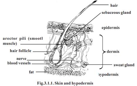
The skin is composed of two major tissues, namely dermis and epidermis. The dermis is mostly formed of connective tissue having fibroblasts, adipose cells and macrophages. It provides the structural strength to the skin. The dermis accommodates nerve endings, hair follicles, smooth muscles and glands.
It is divided into two layers, namely the superficial papillary layer and deeper reticular layer. The papillary layer has projections called papillae. The reticular layer is the major layer of the dermis. It is dense in nature. It is continuous with the hypodermis.
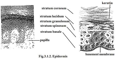
Epidermis: - The epidermis is made up of stratified squamous epithelium. It is separated from the dermis by a basement membrane. It contains melanocytes giving colour to the skin. Many of the cells of the epidermis produce a protein substance called keratin. Hence they are called as keratinocytes.
The deepest layers of the epidermis produce nerve cells by mitosis. As new cells are formed, the older cells are pushed to the surface. The surface cells will protect the inner new cells. Gradually the shape and chemical nature of the surface cells will get altered. Slowly they get filled with keratin.
This process is called keratinization. During this process the epidermis gets divided into five distinct regions or strata .They are the stratum basale, strastratum spinosum, stratum granulosum, strastratum lucidum and stratum Corneum.
Stratum basale is in the deeper region of the epidermis. It consists of one layer of columnar cells. Keratinization of cells begins in this region. Above this layer stratum spinosum is seen. It has 8-10 layers of polygonal cells. The stratum granulosum is the next upper layer. It has 3-5 layers of flattened cells. Above this layer stratum lucidum occurs. It is a thin zone having several layers of dead cells. The top most layers are called the stratum Corneum. It consists of more than 20 layers of dead cells. These cells get filled with keratin. They are said to be cornified. The cornified cells are surrounded by a hard protective envelope.
The skin can be either thick or thin. All five epithelial layers are seen in the thick skin. However stratum Corneum contains more number of cells. Thick skin is formed in the soles of the feet, the palms of hands and tips of fingers. The general body surface has thin skin. In the thin skin each epithelial layer in turn has few layers of cells. There are only one or two layers of cells in stratum granulosum.
Callus: - The regions of skin subjected to constant friction or pressure are thickened to form the callus. The callus has several layers of cells in the stratum Corneum.
Skin colour: - The colour of the skin is due to pigments in the skin. The thickness of the stratum corneum and blood circulation can also cause skin colour. Normally the colour is caused by the pigment melanin. It provides colour to skin, hair and eye. It protects the body from sun’s ultraviolet rays. Melanin is produced by melanocytes. Melanin production is genetically determined. However, hormones and exposure to light can also alter the colour.
Skin derivatives
Hair: - The hairs are integumentary structures. A hair has a root and a shaft. While the shaft projects above the skin, the root remains well below the surface. The base of the root has a hair bulb. It is an expanded region. The shaft and most of the root of the hair are formed of dead keratinized epithelial cells. These are arranged in three concentric layers called the medulla, the cortex and the cuticle. The central axis of the hair is formed of the medulla. Major part of the hair is formed of a single layer of cells.
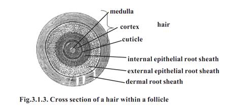
According to the amount and types of melanin, the hair colour may vary. The colour of the hair is genetically determined. During old age the amount of melanin decreases causing white hair. Grey hair has a mixture of faded, unfaded and white hairs.
The hair growth is due to addition of cells at the base of the hair root. The growth stops at specific stages. After a resting period, new hair replaces old hair. The hairs on the head grow for a period of three years and rest for 1-2 years.
The muscle cells found associated with hair follicles is called the arrector pili. Contraction of these muscles cause ‘goose flesh’ making the hairs to ‘stand on end.’
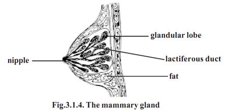
The skin has sebaceous glands and the sweat glands. The sebaceous glands are located in the dermis. They produce an oily substance called the sebum. These glands are connected by a duct to the upper part of the hair follicles. The mammary glands are the modified sweat glands.
The most common type of sweat gland on the skin is the merocrine glands. They are simple coiled tubular glands. They open directly on to the skin through sweat pores. The gland has two parts. They are the deep coiled portion and the duct which passes to the surface of the skin. The number of sweat glands are more in the palms of the hands and soles of the feet.
Nails: - Each nail is made up of two parts. They are the nail root and the nail body. The nail body is the visible part. The nail root is covered by the skin. The proximal and lateral edges of the nail are covered by nail fold.
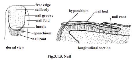
The stratum comeum of the nail fold grows onto the nail body as the eponchium. The free edge of the nail body is the hyponchium. The nail is found placed on the nail matrix and nail bed. A small white region seen at the base of the nail is the lunula. It contains the nail matrix. The nails grow at an average rate of 0.5-1.2 mm per day.
References
T. Sargunam Stephen, Biology (Zoology). First Edition – 2005, Government of Tamilnadu.


|
|
|
|
دراسة: عدم ترتيب الغرفة قد يدل على مشاكل نفسية
|
|
|
|
|
|
|
علماء: تغير المناخ تسبب في ارتفاع الحرارة خلال موسم الحج
|
|
|
|
|
|
|
شعبة فاطمة بنت أسد للدراسات القرآنية تختتم دورة ينابيع الرحمة
|
|
|