


النبات

مواضيع عامة في علم النبات
الجذور - السيقان - الأوراق
النباتات الوعائية واللاوعائية
البذور (مغطاة البذور - عاريات البذور)
الطحالب
النباتات الطبية
الحيوان
مواضيع عامة في علم الحيوان
علم التشريح
التنوع الإحيائي
البايلوجيا الخلوية
الأحياء المجهرية
البكتيريا
الفطريات
الطفيليات
الفايروسات
علم الأمراض
الاورام
الامراض الوراثية
الامراض المناعية
الامراض المدارية
اضطرابات الدورة الدموية
مواضيع عامة في علم الامراض
الحشرات
التقانة الإحيائية
مواضيع عامة في التقانة الإحيائية
التقنية الحيوية المكروبية
التقنية الحيوية والميكروبات
الفعاليات الحيوية
وراثة الاحياء المجهرية
تصنيف الاحياء المجهرية
الاحياء المجهرية في الطبيعة
أيض الاجهاد
التقنية الحيوية والبيئة
التقنية الحيوية والطب
التقنية الحيوية والزراعة
التقنية الحيوية والصناعة
التقنية الحيوية والطاقة
البحار والطحالب الصغيرة
عزل البروتين
هندسة الجينات
التقنية الحياتية النانوية
مفاهيم التقنية الحيوية النانوية
التراكيب النانوية والمجاهر المستخدمة في رؤيتها
تصنيع وتخليق المواد النانوية
تطبيقات التقنية النانوية والحيوية النانوية
الرقائق والمتحسسات الحيوية
المصفوفات المجهرية وحاسوب الدنا
اللقاحات
البيئة والتلوث
علم الأجنة
اعضاء التكاثر وتشكل الاعراس
الاخصاب
التشطر
العصيبة وتشكل الجسيدات
تشكل اللواحق الجنينية
تكون المعيدة وظهور الطبقات الجنينية
مقدمة لعلم الاجنة
الأحياء الجزيئي
مواضيع عامة في الاحياء الجزيئي
علم وظائف الأعضاء
الغدد
مواضيع عامة في الغدد
الغدد الصم و هرموناتها
الجسم تحت السريري
الغدة النخامية
الغدة الكظرية
الغدة التناسلية
الغدة الدرقية والجار الدرقية
الغدة البنكرياسية
الغدة الصنوبرية
مواضيع عامة في علم وظائف الاعضاء
الخلية الحيوانية
الجهاز العصبي
أعضاء الحس
الجهاز العضلي
السوائل الجسمية
الجهاز الدوري والليمف
الجهاز التنفسي
الجهاز الهضمي
الجهاز البولي
المضادات الميكروبية
مواضيع عامة في المضادات الميكروبية
مضادات البكتيريا
مضادات الفطريات
مضادات الطفيليات
مضادات الفايروسات
علم الخلية
الوراثة
الأحياء العامة
المناعة
التحليلات المرضية
الكيمياء الحيوية
مواضيع متنوعة أخرى
الانزيمات

Thoracic Wall
 المؤلف:
Kelly M. Harrell and Ronald Dudek
المؤلف:
Kelly M. Harrell and Ronald Dudek
 المصدر:
Lippincott Illustrated Reviews: Anatomy
المصدر:
Lippincott Illustrated Reviews: Anatomy
 الجزء والصفحة:
 12-7-2021
 9108
9108


+
-
20
Thoracic Wall
The thorax represents the axial region that extends between the neck and the muscular diaphragm. It is characterized by a bony, expandable cage that protects important cardiopulmonary and gastrointestinal (GI) viscera while permitting respiratory movements. The bony thoracic cage is made up of paired ribs (12) that primarily connect the thoracic vertebrae 0 posteriorly with the sternum anteriorly. The thoracic cavity contains the heart, lungs, esophagus, and other associated structures. These con1ent are organized into three main regions : right and left pulmonary cavities and a midline mediastinum. While some structures are self-contained in the thoracic cavity, others pass through the thorax from the neclf to ceacli the abdominal cavity.
The thoracic wall is semi-rigid in its construction, allowing for movement of the ribs with respiration, while also creating a protective cage for thoracic viscera. The inner surface of the wall is lined with a serous parietal pleural layer, which, along with a visceral la ,er, creates two pleural cavities within the thoracic cage.
A. Osteology
The thoracic cage is formeci ya combination of bony and cartilaginous structures, including the sternum, 12 pairs of ribs and costal cartilages, and thoracic vertebrae 1-12 with associated intervertebral discs (Fig. 1).
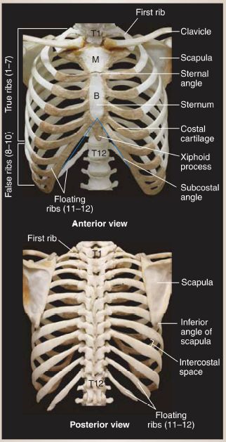
Figure 1: Thoracic cage, bony and cartilaginous components. B = body of sternum, M = manubrium of sternum.
1. Sternum: From superior to inferior, the three parts of the sternum are the manubrium, body, and xiphoid process. The manubrium articulates laterally with the clavicle (sternoclavicular joint) and first costal cartilage and inferiorly with the body. The junction between the manubrium and body is a palpable landmark known as the sternal angle (of Louis) (Fig.2).
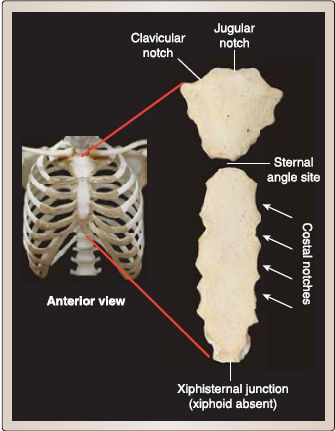
Figure 3.2 : Sternum.
2. Ribs: Ribs are classified as true (1st-7th), false (Bth-10th), or floating (11th-12th), based on direct, indirect, or no articulation by costal cartilage to the sternum, respectively. Floating ribs have no connection to the sternum, as they terminate in the abdominal wall musculature. General rib features include the following (Fig. 3):
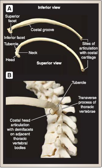
Figure 3 : General rib features. A, Rib parts. B, Costal articulation at thoracic spine.
a. Head: The head articulates with demifacets on the bodies of adjacent thoracic vertebrae. The inferior head facet articulates with the vertebrae that correspond to the rib numerically.
b. Neck: The neck connects head to tubercle region and is not present in 11th-12th ribs.
c. Tubercle: At the transition between neck and body, the tubercle articulates with the corresponding thoracic transverse process and serves as an attachment site for ligamentous support.
d. Body: This thin, flat, long portion of the rib is marked by the costal angle laterally and an internal costal groove that contains intercostal neurovascular bundle. The distal, anterior end of body articulates with costal cartilages, which, in turn, articulate with the sternum.
3. Thoracic vertebrae: Twelve thoracic vertebrae contribute to the posterior boundary of the thoracic cage (see Fig. 3). Thoracic processes serve as attachment sites for ligamentous, muscular, capsular, and costal structures. For a more detailed description of thoracic vertebrae anatomy and clinical considerations.
4. Thoracic apertures: Thoracic apertures mark the superior and inferior boundaries of the thoracic cage. The small, kidney-shaped superior thoracic aperture (thoracic inlet) is bound by the manubrium (anteriorly), 1st rib pair (laterally), and T1 vertebral body (posteriorly). It allows for structural continuity between the neck and thorax. The large, irregularly shaped inferior thoracic aperture is bound by the xiphoid process (anteriorly), costal arch and 12th rib pairs (laterally), and T12 vertebral body (posteriorly). While the muscular diaphragm closes off the inferior thoracic aperture, openings within the diaphragm allow for communication of structures between the thorax and abdomen (Fig. 4).

Figure 4 :Thoracic apertures.
5. Movement: Respiration requires that the thoracic cage be able to expand (increase volume) and retract (decrease volume) to facilitate inspiration and expiration, respectively. Articulations between the vertebrae, ribs, costal cartilages, and sternum, in conjunction with contraction and relaxation of the muscular diaphragm, allow for thoracic cage movement and volume changes in three planes (Fig. 5).
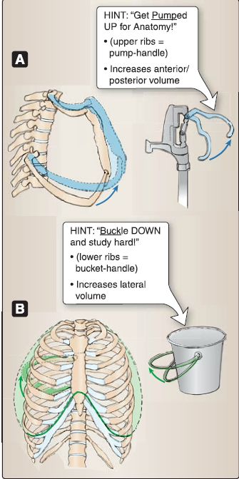
Figure 5: Thoracic cage movements. A, Pumphandle movement. B, Bucket-handle
movement.
a. Anterior/posterior plane: Analogous to a water pump handle, anterior/posterior volume changes involve the upward rotational movement of ribs 1-6 and anterior movement of the sternum.
b. Lateral plane: Like a bucket handle, lateral volume changes involve elevation of the lower ribs, where the lateral portions swing superolaterally.
c. Superior/inferior plane: Superior/inferior volume changes involve contraction and relaxation of the muscular diaphragm, in which contraction flattens the muscle to increase volume and relaxation resumes its dome-shaped appearance.
B. Musculature
The intrinsic muscles of the thoracic wall assist in respiration and are arranged in three layers-superficial, intermediate, and deep. These muscles receive innervation and blood supply from corresponding intercostal nerves (thoracic anterior rami), arteries, and veins (Fig. 6).
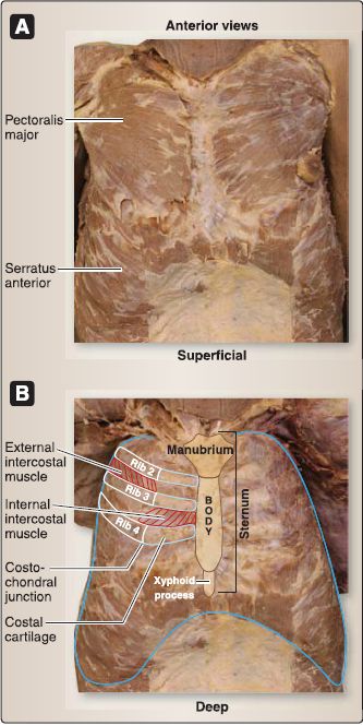
Figure 6 : Anterior thoracic wall musculature. Cadaveric specimens. (Pectoralis major,
pectoralis minor, and serratus anterior muscles reflected bilaterally in B.
1. Superficial layer: External intercostal muscles span the intercostal space from the tubercle to the costochondral junctions, running in an inferomedial direction from superior to inferior ribs. The muscle is replaced anteriorly by external intercostal membranes along the costal cartilage to the sternum.
2. Intermediate layer: Internal intercostal muscles span the intercostal space from the sternum and costal cartilages posteriorly to the angle of the ribs, running in an inferolateral direction from superior to inferior ribs. The muscle is replaced posteriorly by internal intercostal membranes.
3. Deep layer: Innermost intercostal and transversus thoracis muscles represent this incomplete layer. Innermost intercostal muscles are most developed posterolaterally and may span one or two intercostal spaces. Those muscles spanning two intercostal spaces are called subcostal muscles. Transversus thoracis muscles consist of four to five paired slips of muscle that attach between the posterior surface of the inferior sternum to superior costal cartilages.
4. Diaphragm: Although not an intrinsic muscle of the thoracic wall, the diaphragm is the most important respiratory muscle. This dome-shaped muscle arises from the costal margins of the inferior thoracic aperture and upper lumbar vertebrae. The strong central tendon of the diaphragm serves as a common insertion point for the peripheral fibers. With inspiration, the diaphragm flattens and then assumes its relaxed dome shape during expiration. The diaphragm has three posterior openings to allow for passage of the inferior vena cava (IVC), esophagus, and aorta at vertebral levels T8, T10, and T12, respectively.
5. Accessory respiratory muscles: Muscles that contribute to the thoracic wall and aid respiratory function include serratus posterior superior, serratus posterior inferior, and levator costarum. These muscles may also play an important role in proprioception of the thoracic cage.
6. Axioappendicular, neck, and abdominal muscles: Muscles that have attachments to the ribs, sternum, and costal cartilages also contribute to the structure and stability of the thoracic cage. These muscles include pectoralis major, pectoralis minor, subclavius, scalenes, serratus anterior, external abdominal oblique, internal abdominal oblique, transversus abdominis, and rectus abdominis.
C. Innervation
Thoracic spinal nerves 1-11 give rise to 11 paired thoracic anterior rami known as intercostal nerves. The T12 anterior rami pair travel inferior to the 12th rib and are known as subcostal nerves. At the angle of the rib, a typical intercostal nerve travels between the innermost and internal intercostal muscles along the costal groove with intercostal veins and arteries. The arrangement of the intercostal neurovascular bundle is such that, from superior to inferior, their order is vein, artery, and nerve (mnemonic = VAN) (Fig. 7).
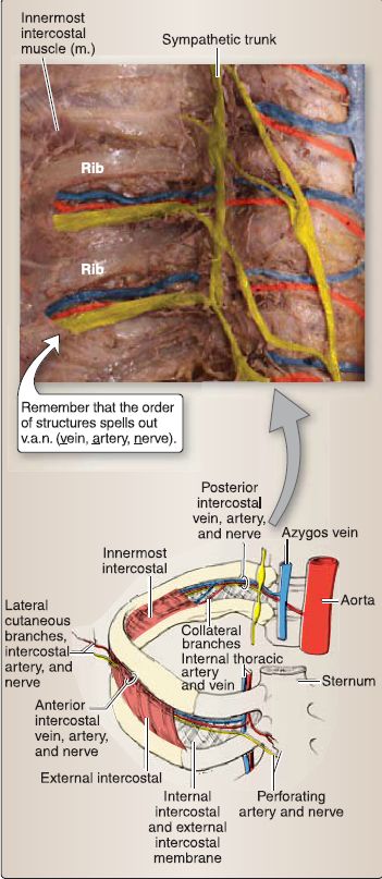
Figure 7 : lntercostal neuromuscular bundle. lntercostal space showing bundle distribution (bottom). Posterior thoracic wall showing bundle orientation and path (top).
1. lntercostal nerves: These nerves provide innervation to the skin and muscle of the thoracic wall. Along their course, intercostal nerves give off lateral and anterior cutaneous branches to the areas of skin that correspond to thoracic dermatomes T1-T11 . As a result of upper limb development, T1 and T2 spinal nerve dermatome distribution is also found along the medial arm and forearm.
2. Muscular and sensory branches: Muscular branches arise along the course of the intercostal nerves to innervate the muscles of the thoracic wall. Sensory branches also arise to mediate sensation from peripheral portions of the diaphragm and diaphragmatic and costal pleura. The diaphragm receives motor innervation from the phrenic nerve (C3-C5).
D. Vasculature
lntercostal arteries arise primarily from either the thoracic aorta as posterior intercostal arteries or the internal thoracic (internal mammary) arteries as anterior intercostal arteries. Posterior and anterior intercostal arteries anastomose with each other. Each of the 11 intercostal spaces receives blood supply from posterior and anterior intercostal arteries and collateral branches that arise from posterior intercostal arteries. The subcostal arteries supply a small area inferior to the 12th rib (Fig. 8). Eleven pairs of posterior and anterior intercostal veins drain the thoracic wall primarily by way of the azygos venous system or internal thoracic veins, respectively. Subcostal veins drain a small area inferior to the 12th rib.

Figure 8 : lntercostal blood supply. A, Arterial distribution. B, Venous distribution.
 الاكثر قراءة في علم التشريح
 اخر الاخبار
اخبار العتبة العباسية المقدسة

الآخبار الصحية


 قسم الشؤون الفكرية يصدر كتاباً يوثق تاريخ السدانة في العتبة العباسية المقدسة
 "المهمة".. إصدار قصصي يوثّق القصص الفائزة في مسابقة فتوى الدفاع المقدسة للقصة القصيرة
 (نوافذ).. إصدار أدبي يوثق القصص الفائزة في مسابقة الإمام العسكري (عليه السلام)


