

 النبات
 الحيوان
 الأحياء المجهرية
 علم الأمراض
 التقانة الإحيائية
 التقنية الحيوية المكروبية
 التقنية الحياتية النانوية
 علم الأجنة
 الأحياء الجزيئي
 علم وظائف الأعضاء
 الغدد
الغدد
 المضادات الحيوية
|
Read More
Date: 12-7-2021
Date: 5-11-2015
Date: 5-11-2015
|
Posterior Abdominal Wall
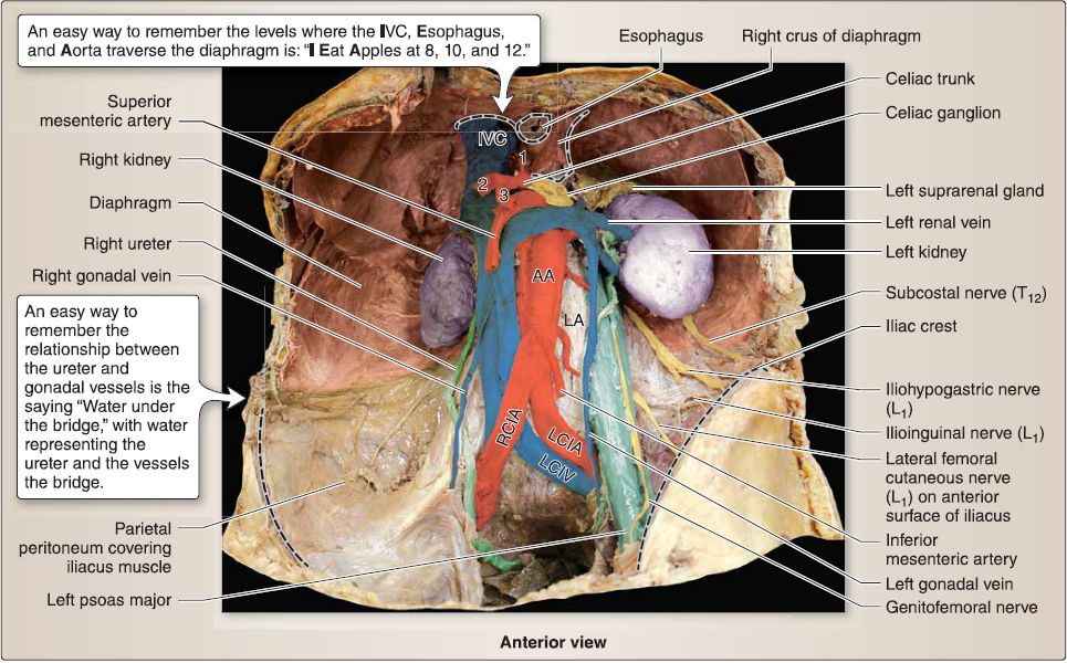
The posterior abdominal wall comprises structures that lie posterior to the parietal peritoneum in the abdominal cavity (Fig. 1). This region is bound posteriorly by lumbar vertebrae (L 1-L5) and intervertebral discs; superiorly by portions of the muscular diaphragm; posterolaterally by psoas major, quadratus lumborum, iliacus, and transversus abdominus; and anteriorly and inferiorly by posterior parietal peritoneum. In addition to the structures that bind this region, it contains the kidneys, suprarenal glands, ureters, abdominal aorta and branches, IVC and tributaries, sympathetic trunks, lymph nodes, lumbar plexus branches, and variable amounts of fat.

Figure 1: Posterior abdominal wall structure. Gastrointestinal viscera and peritoneum removed to reveal posterior abdominal wall structures. 1 = left gastric artery, 2 = common hepatic artery, 3 = splenic artery, AA = abdominal aorta, IVG = inferior vena cava, LA = lumbar artery, LCIA = left common iliac artery, LCIV = left common iliac vein, RCIA = right common iliac artery.
A. Viscera
Viscera contained in the posterior abdominal wall region include the right and left kidneys, ureters, and suprarenal glands.
B. Muscles
The muscles that support the posterior abdominal wall act on both the spine and the lower limbs (thighs). Some of these muscles also contribute to the formation of the anterolateral abdominal wall. Endoabdominal fascia lies between the muscles and posterior parietal peritoneum, which is continuous laterally with the transversalis fascia of the anterolateral abdominal wall (Fig. 2).

Figure 2 :Posterior abdominal wall muscles and nerves. Left posterior abdominal
wall with left kidney reflected to reveal musculature and nerves.
1. Psoas major: This muscle originates on lumbar vertebrae transverse processes, bodies (f12 as well), and associated intervertebral discs and inserts with the iliacus onto the lesser trochanter of the femur. This muscle is a powerful flexor of the thigh and spine and also laterally flexes the lumbar spine. It is innervated by anterior rami of L1-L3 . The superior portion of psoas fascia thickens to form the medial arcuate ligament.
2. Quadratus lumborum: This muscle originates from lumbar transverse processes and the lower border of rib 12, medially, and inserts onto the iliac crest and iliolumbar ligaments. It extends and laterally flexes the lumbar spine and stabilizes the 12th rib during inspiration. It receives innervation from anterior rami of T12 and L1-L4. The superior portion of quadratus lumborum fascia (anterior layer of thoracolumbar fascia) thickens to form the lateral arcuate ligament.
3. lliacus: This muscle originates from the iliac fossa and inserts with psoas major onto the lesser trochanter of the femur. It flexes the thigh and stabilizes the hip joint and is innervated in the pelvis by the femoral nerve.
4. Transversus abdominis: The deepest of the flat lateral abdominal wall muscles, transversus abdominis originates posteriorly and is located internally, just lateral to quadratus lumborum and inferior to the posterior diaphragm. See I1.A.1.c. for more details.
5. Diaphragm: The diaphragm separates the thoracic and abdominal cavities and functions in respiration. Centrally, the diaphragm is characterized by an aponeurotic central tendon. Along its periphery the diaphragm becomes more muscular. The muscular arrangement and attachments of the lumbar portion of the diaphragm create a series of hiatuses to permit passage of vessels and viscera between thoracic and abdominal cavities.
a. Attachments: The muscular portion of the diaphragm is attached along the inner boundary of the thoracic outlet (sternal and costal portions) and on the first through third lumbar vertebrae {lumbar portion). Posteriorly, the lumbar portion divides around the vertebral bodies, forming two extensions of muscle-right and left crura (crus means "leg"). These crura serve to anchor the diaphragm posteriorly. In the midline, the left and right crura are joined by the median arcuate ligament, a fibrous structure that completes the opening for the aorta.
b. Hiatuses: The diaphragm has three main hiatuses to allow for the IVC, esophagus, and descending aorta to pass between the thoracic and abdominal cavities. Each passageway occurs at a specific vertebral level.
[1] TB, caval opening (for IVC): Located in the central tendon region, terminal branches of the right phrenic nerve and lymphatic vessels also pass through this opening.
[2] T10, esophageal hiatus: This opening is located in the right crus of the diaphragm. Hiatal fibers constrict the esophagus during contraction of the diaphragm, acting as a sphincter. It also transmits vagal trunks, esophageal vessels {left gastric branches), and lymphatic vessels.
[3] T12, aortic hiatus: This opening is located between the left and right crura of the diaphragm, and diaphragm movement does not interfere with blood flow through the aorta. The aponeurotic arch between right and left crura- median arcuate ligament-caps the aorta in the midline.
c. Neurovascular and lymphatic supply: The inferior {abdominal) surface of the diaphragm receives blood supply from the inferior phrenic vessels and innervation from phrenic nerves (G3-G5) and intercostal nerves {laterally). Lymph from the inferior surface of the diaphragm drains into phrenic, anterior diaphragmatic, and lumbar lymph nodes.
C. Blood supply
blood supply to the posterior abdominal wall is achieved through paired branches of the abdominal aorta (inferior phrenic, lumbar, and subcostal arteries) and tributaries of the IVG and azygos system (posterior intercostal and lumbar veins).
D. Innervation
Innervation of the posterior abdominal wall musculature is described above. Visceral innervation of the kidneys, suprarenal glands. Autonomic plexuses associated with the aorta are also found in the posterior abdominal wall region, some extending to viscera in this region as well. Branches of the lumbar plexus, along with the subcostal nerve (T12), course along posterior abdominal wall musculature to provide motor and sensory innervation to abdominal wall and lower limb structures (see Fig. 2).
1. Subcostal (T12): This branch travels anterior to the transversus abdominis before piercing the muscle to run between it and theinternal oblique to innervate the external oblique and overlying anterolateral abdominal wall skin.
2. lliohypogastric (L1): This branch travels anterior to the quadratus lumborum, then between the transversus abdominis and internal oblique to innervate muscles and skin of the anterolateral abdominal wall. It may arise from a common trunk with ilioinguinal nerve.
3. llioinguinal (L1): This branch travels anterior to quadratus lumborum, then between the transversus abdominis and internal oblique to innervate muscles and skin of the anterolateral abdominal wall. It may arise from a common trunk with iliohypogastric nerve. The ilioinguinal nerve travels through portions of the inguinal canal to reach the inferior portion of this region.
4. Lateral femoral cutaneous (L2-L:J: This branch travels anterior to iliacus, then deep to the inguinal ligament just medial to the anterior superior iliac spine to innervate skin on the superolateral thigh.
5. Femoral (L2-L4): This branch travels deep to psoas major before exiting laterally, passing deep to the inguinal ligament to enter the femoral triangle. The femoral nerve provides motor and sensory innervation to anterior thigh and medial leg/foot structures.
6. Genitofemoral (L1-LJ: This branch travels on the anterior surface of psoas major and splits distally into a genital branch and femoral branch, which innervate structures of the perineum and anterior thigh, respectively.
7. Obturator (L2-L4): This branch travels deep to psoas major before exiting medially, traveling through a small opening in the obturator fascia to reach and innervate structures in the medial thigh.
E. Lymphatics
Lymphatic structures are intimately related to the abdominal aorta . These centrally located nodes and vessels collect lymph from peripheral viscera and the posterior abdominal wall structures. Specifically, lymph from the posterior abdominal wall drains initially into right Interpretation of abdominal imaging requires a thorough approach. A solid understanding of the abdominal anatomy and spatial relationships is very important and useful in both plain film and computed tomography (CT) interpretation.


|
|
|
|
إدارة الغذاء والدواء الأميركية تقرّ عقارا جديدا للألزهايمر
|
|
|
|
|
|
|
شراء وقود الطائرات المستدام.. "الدفع" من جيب المسافر
|
|
|
|
|
|
|
العتبة العبّاسيّة: البحوث الّتي نوقشت في أسبوع الإمامة استطاعت أن تثري المشهد الثّقافي
|
|
|