آخر المواضيع المضافة

 النبات
الحيوان
الأحياء المجهرية
علم الأمراض
التقانة الإحيائية
التقنية الحيوية المكروبية
التقنية الحياتية النانوية
علم الأجنة
الأحياء الجزيئي
علم وظائف الأعضاء
الغدد
المضادات الحيوية
النبات
الحيوان
الأحياء المجهرية
علم الأمراض
التقانة الإحيائية
التقنية الحيوية المكروبية
التقنية الحياتية النانوية
علم الأجنة
الأحياء الجزيئي
علم وظائف الأعضاء
الغدد
المضادات الحيوية| Muscular System |


|
|
Read More
Date: 10-7-2021
Date: 16-7-2021
Date: 1-8-2021
|
Muscular System
Locomotion and bodily movements are characteristic features of the animals. The movements are effected by various cell organelles such as cilia, flagella and organs like muscles. Muscular movements are more powerful and energetic. The skeletal muscles apart from their role in smarter movements provide beautiful shapes to the body. The inner smooth muscles of the visceral organs make them work like machines all through the life period. The muscle cells function like small motors to produce the forces responsible for the movement of the arms, legs, heart and other part of the body. Thus the highly specialized muscle tissues are responsible for the mechanical processes in the body.
Based on structure, functioning and occurrence three different types of muscle tissues have been identified. They are the skeletal, visceral and cardiac muscles.
1-Skeletal muscles or striped muscles: These muscles are attached to the bones. The muscle cells are long and cylindrical. These voluntary muscles cause body movements.
2-Visceral muscles or nonstriated muscles: These are found in the walls of the inner organs such as blood vessels, stomach and intestine. The muscle cells are spindle shaped. These are involuntary in nature.
3-Cardiac muscle: These are found in the wall of the heart. The muscle cells are cylindrical and branched. The muscles are involuntary in nature.
Skeletal muscles
The skeletal muscles are attached to bones by tendons. The tendons help to transfer the forces developed by skeletal muscles to the bones. These muscles are covered by sheets of connective tissue called fascia.
Tendons: These are connective tissue structures showing slight elasticity. They are like cords or straps strongly attached to bones. The tensile strength of tendons is nearly half that of steel. A tendon having 10 mm diameter can support 600 - 1000 kg.
Fascia: These are assemblages of connective tissues lining skeletal muscles as membranous sheets. The fascia may be superficial or deep. The superficial fascia is a layer of loose connective tissue found in between skin and muscles. The deep fascia is collagen fibres found as a tough inelastic sheath around the musculature. They run between groups of muscles and connect with the bones.
Shapes of muscles
There is a wide variety of shapes and sizes in muscles. Based on general shape and the orientation or muscle fibres in relation to the direction of pull, they can be grouped into two classes.
1-Parallel: These muscle fibres are parallel to the line of pull. The muscles may be flat, short, quadrilateral or long and strap like. The individual fibres run the entire length of the muscle.
2-Oblique: These muscle fibres are oblique to the line of pull. The muscles may be triangular or pennate (feather-like). The pennate forms may be unipennate, bipennate, multipennate or circumpennate. Some muscles have a spiral or twisted arrangement.
Naming of muscles.
The muscles are named according to their size, shape, position and
action.
Shape Size
Deltoid - triangular major - large
Quadratus - square minor - small
Gracilis - slender longus - long
lattismus - broadest
Number of Heads Position
biceps - 2 heads dorsi - of the back
triceps - 3 heads pectoralis - of the chest
quandriceps - 4 heads brachii - of the arm
anterior, posterior
Depth Action
superficialis - superficial extensor
internus - internal, flexor constrictor
profundus - deep Flexor
Distribution of muscles
I. Muscles of the head
There are two groups of muscles. They are craniofacial and masticatory muscles. The craniofacial muscles are related to eye orbital margins, eyelids, nose, nostrils, lips, cheeks, mouth, pinna, and the scalp. These muscles are also known as muscles of facial expression. Among these muscles those that are related to the lip movement are significant. The facial expression is mostly due to lip movement and positioning of the lips. Such thought related movements are caused by several muscles associated with lips and the skin around the mouth. Since orbicularis oris and buccinators muscles provide lip movement for kissing posture they are known as “kissing muscles”. Smiling is accomplished by zygomasticus major and minor, levator anguli oris and risorius. The muscles of the lips can also provide expressions such as sneering and frowning. The chin dimples are located between the mentalis muscles.
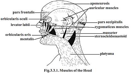
The masticatory (or speech) muscles move the mandible of the lower jaw. The muscles responsible for this movement are masseter temporalis and pterygoid. Tongue movements are caused by intrinsic and extrinsic muscles. Swallowing of food is facilitated by several muscles related to the mouth, roof of pharynx, uvula and other regions.
II. Muscles of the Neck region
The movements of the neck region are caused by cervical, suprahyoid, infrahyoid and vertebral muscles.
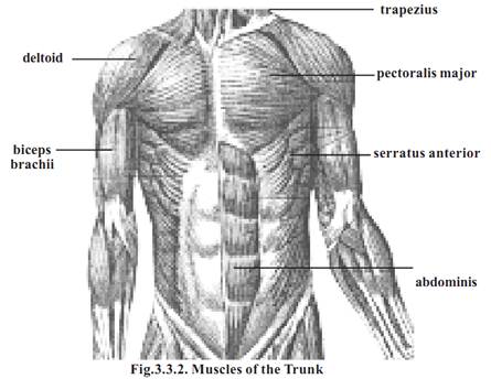
III. Muscles of the Trunk region
The muscles of the vertebral column help to bend and rotate the body. These are strong back muscles that help the trunk to maintain erect posture. The most prominent muscles of this region are the erector spinae, longissimus and spinalis.
Four important thoracic muscle groups are associated with the process of breathing. While the process of inspiration is due to scalene and external intercostal muscles, the expiration is performed due to internal intercostals and transverse thoraces. Major breathing movement is due to diaphragm, a curved musculofibrous sheet that separates thoracic and abdominal cavities.
Abdominal muscles can aid in forced expiration, vomiting, defaecation, urination and childbirth.
The inferior opening of the pelvic bone is covered by pelvic diaphragm muscles. Below these muscles perineum is present. The perineum and other “subfloor” muscles form the urogenital diaphragm. Pelvic and urogenital diaphragm may get stretched in pregnancy due to weight of the foetus. However by specific exercises they can be strengthened.
IV. Muscles of the upper limb
The hands are attached to the pectoral girdle and to the vertebral column by large conspicuous muscles such as trapezius, rhomboid major and minor, levator scapulae and lattissimus dorsi.
The trapezius is a flat, triangular muscle. It extends over the back of the neck and upper thorax. It maintains the level and poise of the shoulder. It helps to rotate the scapula forward, so that the arm can be raised above the head. It helps to bend the neck backwards and laterally.
Latissimus dorsi is a large flat triangular muscle. It is a conspicuous muscle stretching over the lumbar region and lower thorax. This muscle is useful in adduction, extension and medial rotation of the humerus. It helps in the backward swinging of the arm. By raising the arm above the head it helps to pull the trunk upwards and forwards. It is useful in violent expiratory activities such as coughing or sneezing. It helps in deep inspiration.
Serratus anterior and pectoralis major connect the ribs to the scapula. Pectoralis major extends from the upper thorax and abdomen to act on the humerus. It is a fan shaped muscle. It spreads between the clavicle and the 7th costal cartilage in the front of the chest. It helps to swing the extended arm forward and medially. It helps in climbing. It is active in deep inspiration.
The muscles of the upper arm are the Coracobrachialis, biceps, triceps and brachialis. The coracobrachialis arises from the coracoid bone in the shoulder and ends in the humerus of the upper arm. It helps to move the arm forward and medially. The biceps brachii is a large fusiform muscle. It has two proximal heads for attachment. They are connected to the coracoid and shoulder joint. The lower head ends in the radius of the lower arm. It is a powerful muscle causing flexing of the hand. The triceps arises by three heads from scapula and upper part of humerus on the posterior side. The wrist, hand and finger movements are caused by several extrinsic and intrinsic hand muscles. A detailed study of them could be made in higher classes.
V. Muscles of the lower limb
Thigh movements are caused by anterior, postereolateral and deep muscles. The anterior muscles are the iliacus and psoas major which help to flex the thigh. The gluteus maximus form the mass of the buttocks region. Leg movement is caused by the anterior thigh muscles, quadriceps femoris and sartorius. The sartorius is the longest muscle of the body. It runs from the hip to the knee. Muscle movement of ankle foot and toe are caused by several groups of extrinsic and intrinsic muscles. A detailed study of them could be made in higher classes.
References
T. Sargunam Stephen, Biology (Zoology). First Edition – 2005, Government of Tamilnadu.


|
|
|
|
التوتر والسرطان.. علماء يحذرون من "صلة خطيرة"
|
|
|
|
|
|
|
مرآة السيارة: مدى دقة عكسها للصورة الصحيحة
|
|
|
|
|
|
|
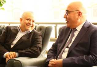
نحو شراكة وطنية متكاملة.. الأمين العام للعتبة الحسينية يبحث مع وكيل وزارة الخارجية آفاق التعاون المؤسسي
|
|
|