

 النبات
 الحيوان
 الأحياء المجهرية
 علم الأمراض
 التقانة الإحيائية
 التقنية الحيوية المكروبية
 التقنية الحياتية النانوية
 علم الأجنة
 الأحياء الجزيئي
 علم وظائف الأعضاء
 الغدد
الغدد
 المضادات الحيوية
|
Read More
Date: 25-7-2016
Date: 3-8-2016
Date: 8-1-2017
|
Lingual Tonsil
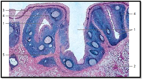
The root of the tongue between sulcus terminalis and epiglottis features tonsillar crypts 1 . These are short narrow caverns (invaginations). The tonsillar crypts may continue in the secretory ducts of mucous glands 2 or have a blind end. The crypts are lined by multilayered nonkeratinizing squamous epithelium and surrounded by lymphatic tissue. The figure shows the multilayered squamous epithelium 3 , which covers the root of the tongue and its crypts (invaginations, caverns) 1 . The lymphoreticular tissue (stained deep blue) 4 underneath the epithelium is part of the lamina propria. Numerous more lightly stained areas are found in the lymphoreticular tissue. These are secondary follicles. The lymphoreticular tissue is separate d from the surrounding tissue by a more or less complete connective tissue capsule 5 .
1 Tonsillar crypts
2 Mucous glands of the root of the tongue, glandulae linguales posteriores
3 Epithelium of the lingual mucous membrane
4 Lymphoreticular tissue with germinal center s
5 Connective tissue capsule
Stain: alum hematoxylin-eosin; magnification: × 14

Lingual Tonsil
This vertical section through the root of the tongue shows the lingual follicles 1. The top part of the figure reveals the multilayered nonkeratinizing squamous epithelium of the root of the tongue 2 and the underlying lymphoreticular tissue 1 . Striate d muscle fibers of the lingual musculature 4 are visible in the lower half of the image. The muscle cells are interspersed with the lobules of the posterior lingual mucous glands 5 . The connective tissue is stained blue.
1 Lymphoreticular tissue
2 Epithelium of the root of the tongue
3Crypt
4 Tongue musculature
5 Mucous glands
Stain: azan; magnification: × 12

References
Kuehnel, W.(2003). Color Atlas of Cytology, Histology, and Microscopic Anatomy. 4th edition . Institute of Anatomy Universitätzu Luebeck Luebeck, Germany . Thieme Stuttgart · New York .


|
|
|
|
تفوقت في الاختبار على الجميع.. فاكهة "خارقة" في عالم التغذية
|
|
|
|
|
|
|
أمين عام أوبك: النفط الخام والغاز الطبيعي "هبة من الله"
|
|
|
|
|
|
|
قسم شؤون المعارف ينظم دورة عن آليات عمل الفهارس الفنية للموسوعات والكتب لملاكاته
|
|
|