آخر المواضيع المضافة

 النبات
الحيوان
الأحياء المجهرية
علم الأمراض
التقانة الإحيائية
التقنية الحيوية المكروبية
التقنية الحياتية النانوية
علم الأجنة
الأحياء الجزيئي
علم وظائف الأعضاء
الغدد
المضادات الحيوية
النبات
الحيوان
الأحياء المجهرية
علم الأمراض
التقانة الإحيائية
التقنية الحيوية المكروبية
التقنية الحياتية النانوية
علم الأجنة
الأحياء الجزيئي
علم وظائف الأعضاء
الغدد
المضادات الحيوية| Artery and Vein |


|
|
Read More
Date: 19-1-2017
Date: 25-7-2016
Date: 26-7-2016
|
Artery and Vein
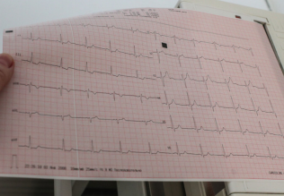
This preparation is from the submucosal tissue of the stomach. The micrograph shows cross-sections of a small artery (bottom right) and a vein. The epithelial cell nuclei of the tunica intima protrude into the vascular lumen and make the opening look like an eyelet 1 . Connective tissue dominates in the tunica media of the venous wall. In contrast, in the tunica me dia of the arterial wall circular smooth muscle bundles 2 are more prevalent. Folds of the tunica intima form the venous valves 3 , which extend into the venous lumen.
1 Endothelial cell nuclei
2 Circular musculature
3 Venous valve
4 Arteriole (longitudinal cut)
5 Arteriole (cut across the axis)
6 Connective tissue from the submucous coat (stomach)
Stain: alum hematoxylin-eosin; magnification: × 300

References
Kuehnel, W.(2003). Color Atlas of Cytology, Histology, and Microscopic Anatomy. 4th edition . Institute of Anatomy Universitätzu Luebeck Luebeck, Germany . Thieme Stuttgart · New York .


|
|
|
|
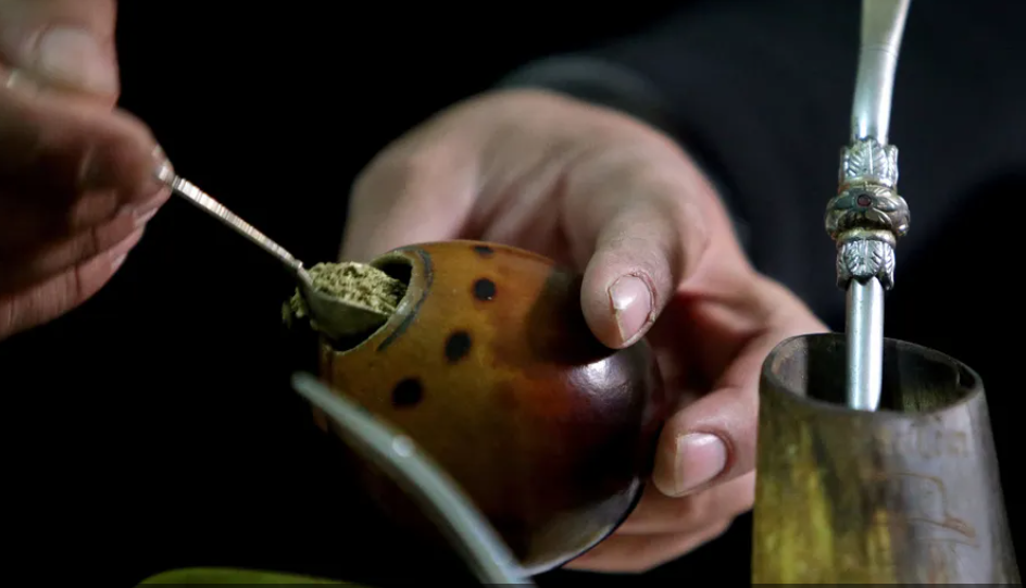
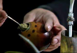
"إنقاص الوزن".. مشروب تقليدي قد يتفوق على حقن "أوزيمبيك"
|
|
|
|
|
|
|
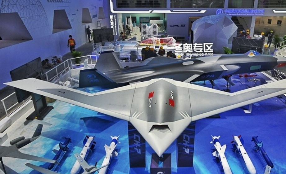
الصين تحقق اختراقا بطائرة مسيرة مزودة بالذكاء الاصطناعي
|
|
|
|
|
|
|
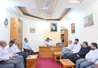
قسم شؤون المعارف ووفد من جامعة البصرة يبحثان سبل تعزيز التعاون المشترك
|
|
|