آخر المواضيع المضافة

 النبات
الحيوان
الأحياء المجهرية
علم الأمراض
التقانة الإحيائية
التقنية الحيوية المكروبية
التقنية الحياتية النانوية
علم الأجنة
الأحياء الجزيئي
علم وظائف الأعضاء
الغدد
المضادات الحيوية
النبات
الحيوان
الأحياء المجهرية
علم الأمراض
التقانة الإحيائية
التقنية الحيوية المكروبية
التقنية الحياتية النانوية
علم الأجنة
الأحياء الجزيئي
علم وظائف الأعضاء
الغدد
المضادات الحيوية| Microvilli-Microplicae |


|
|
Read More
Date: 8-1-2017
Date: 8-1-2017
Date: 19-1-2017
|
Microvilli-Microplicae
The cell surface can also be enlarged by raised, leaf-like formations of the plasmalemma—i.e., by outward extension in the form of folds. The figure shows flat epithelial cells from the canine tongue with a dense pattern of microplicae —i.e., raise d folds of the plasma membrane. Such microplicae folds are commonly observe d in a multitude of patterns. Microplicae also exist at the bottom face of flat epithelial cells, and therefore may play a role in intercellular adhesion. The more pronounced “edges” in this figure correspond to the cell borders. In transmission electron microscopy, vertical cuts show microplicae of ten as short, stump-shape d microvilli. Located on top of the epithelial cells are two erythrocytes and a rod-shaped form, which can be categorize d as an oral cavity saprophyte.
Scanning electron microscopy; magnification: × 3600

References
Kuehnel, W.(2003). Color Atlas of Cytology, Histology, and Microscopic Anatomy. 4th edition . Institute of Anatomy Universitätzu Luebeck Luebeck, Germany . Thieme Stuttgar t · New York .


|
|
|
|
اكتشاف تأثير صحي مزدوج لتلوث الهواء على البالغين في منتصف العمر
|
|
|
|
|
|
|
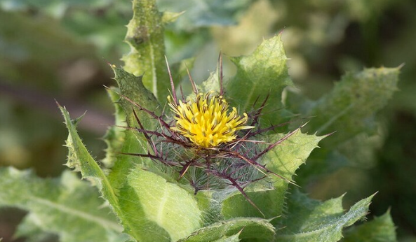
زهور برية شائعة لتر ميم الأعصاب التالفة
|
|
|
|
|
|
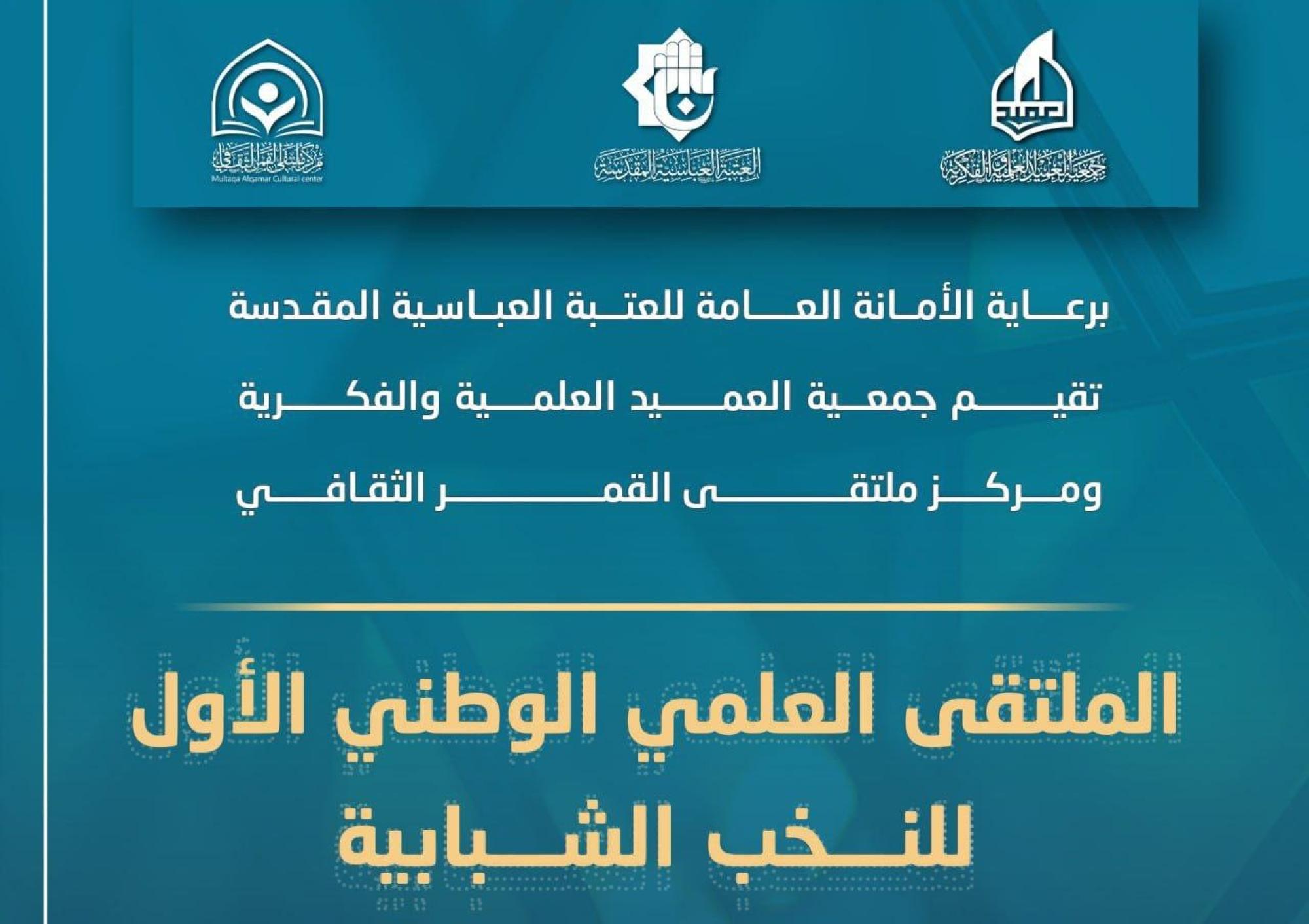
جمعيّة العميد وقسم الشؤون الفكريّة تدعوان الباحثين للمشاركة في الملتقى العلمي الوطني الأوّل
|
|
|
|
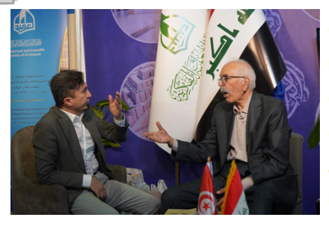
الأمين العام المساعد لجامعة الدول العربية السابق: جناح جمعية العميد في معرض تونس ثمين بإصداراته
|
|
|
|
المجمع العلمي يستأنف فعاليات محفل منابر النور في واسط
|
|
|
|
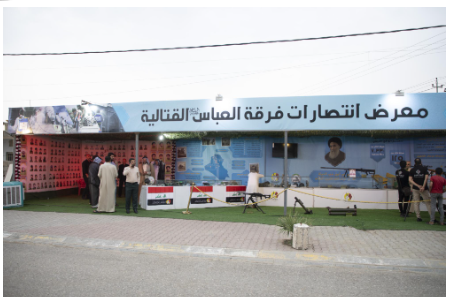
برعاية العتبة العباسيّة المقدّسة فرقة العبّاس (عليه السلام) تُقيم معرضًا يوثّق انتصاراتها في قرية البشير بمحافظة كركوك
|