آخر المواضيع المضافة

 النبات
الحيوان
الأحياء المجهرية
علم الأمراض
التقانة الإحيائية
التقنية الحيوية المكروبية
التقنية الحياتية النانوية
علم الأجنة
الأحياء الجزيئي
علم وظائف الأعضاء
الغدد
المضادات الحيوية
النبات
الحيوان
الأحياء المجهرية
علم الأمراض
التقانة الإحيائية
التقنية الحيوية المكروبية
التقنية الحياتية النانوية
علم الأجنة
الأحياء الجزيئي
علم وظائف الأعضاء
الغدد
المضادات الحيوية| Wound Healing |


|
|
Read More
Date: 25-2-2016
Date: 26-2-2016
Date: 28-2-2016
|
Wound Healing
The two processes of healing, can occur during healing of a diseased organ or during healing of a wound. A wound can be accidental or surgical. Now, we will discuss skin wound healing to demonstrate the two basic processes of healing.
Healing of a wound demonstrates both epithelial regeneration (healing of the epidermis) and repair by scarring (healing of the dermis).
There are two patterns of wound healing depending on the amount of tissue damage:
1. Healing by first intention (Primary union)
2. Healing by second intention
These two patterns are essentially the same process varying only in amount.
1. Healing by first intention (primary union)
The least complicated example of wound healing is the healing of a clean surgical incision. The wound edges are approximated by surgical sutures, and healing occurs with a minimal loss of tissue. Such healing is referred to, surgically, as “primary union” or “healing by first intention”. The incision causes the death of a limited number of epithelial cells as well as of dermal adnexa and connective tissue cells; the incisional space is narrow and immediately fills with clotted blood, containing fibrin and blood cells; dehydration of the surface clot forms the well-known scab that covers the wound and seals it from the environment almost at once.
Within 24 hours, neutrophils appear at the margins of the incision, moving toward the fibrin clot. The epidermis at its cut edges thickens as a result of mitotic activity of basal cells and, within 24 to 48 hours, spurs of epithelial cells from the edges both migrate and grow along the cut margins of the dermis and beneath the surface scab to fuse in the midline, thus producing a continuous but thin epithelial layer.
By day 3, the neutrophils have been largely replaced by macrophages. Granulation tissue progressively invades the incisional space. Collagen fibers are now present in the margins of the incision, but at first these are vertically oriented and do not bridge the incision. Epithelial cell proliferation continues, thickening the epidermal covering layer. By day 5, the incisional space is filled with granulation tissue. Neovascularization is maximal. Collagen fibrils become more abundant and begin to bridge the incision. The epidermis recovers its normal thickness and differentiation of surface cells yields a mature epidermal architecture with surface keratinization.
During the second week, there is continued accumulation of collagen and proliferation of fibroblasts. Leukocytic infiltrate, edema, and increased vascularity have largely disappeared. At this time, the long process of blanching begins, accomplished by the increased accumulation of collagen within the incisional scar, accompanied by regression of vascular channels.
By the end of the first month, the scar comprises a cellular connective tissue devoid of inflammatory infiltrate, covered now by an intact epidermis. The dermal appendages that have been destroyed in the line of the incision are permanently lost. Tensile strength of the wound increases thereafter, but it may take months for the wounded area to obtain its maximal strength.
2. Healing by second intention (secondary union)
When there is more extensive loss of cells and tissue, such as occurs in infarction, inflammatory ulceration, abscess formation, and surface wounds that create large defects, the reparative process is more complicated. The common denominator in all these situations is a large tissue defect that must be filled. Regeneration of parenchymal cells cannot completely reconstitute the original architecture. Abundant granulation tissue grows in from the margin to complete the repair. This form of healing is referred to as “secondary union” or “healing by second intention.”
Secondary healing differs from primary healing in several respects:
1. Inevitably, large tissue defects initially have more fibrin and more necrotic debris and exudate that must be removed. Consequently, the inflammatory reaction is more intense.
2. Much larger amounts of granulation tissue are formed. When a large defect occurs in deeper tissues, such as in a viscus, granulation tissue bears the full responsibility for its closure, because drainage to the surface cannot occur.
3. Perhaps the feature that most clearly differentiates primary from secondary healing is the phenomenon of wound contraction, which occurs in large surface wounds.
4. Healing by second intention takes much longer than when it occurs by first intention.
References
Bezabeh ,M. ; Tesfaye,A.; Ergicho, B.; Erke, M.; Mengistu, S. and Bedane,A.; Desta, A.(2004). General Pathology. Jimma University, Gondar University Haramaya University, Dedub University.


|
|
|
|
اكتشاف تأثير صحي مزدوج لتلوث الهواء على البالغين في منتصف العمر
|
|
|
|
|
|
|
زهور برية شائعة لتر ميم الأعصاب التالفة
|
|
|
|
|
|
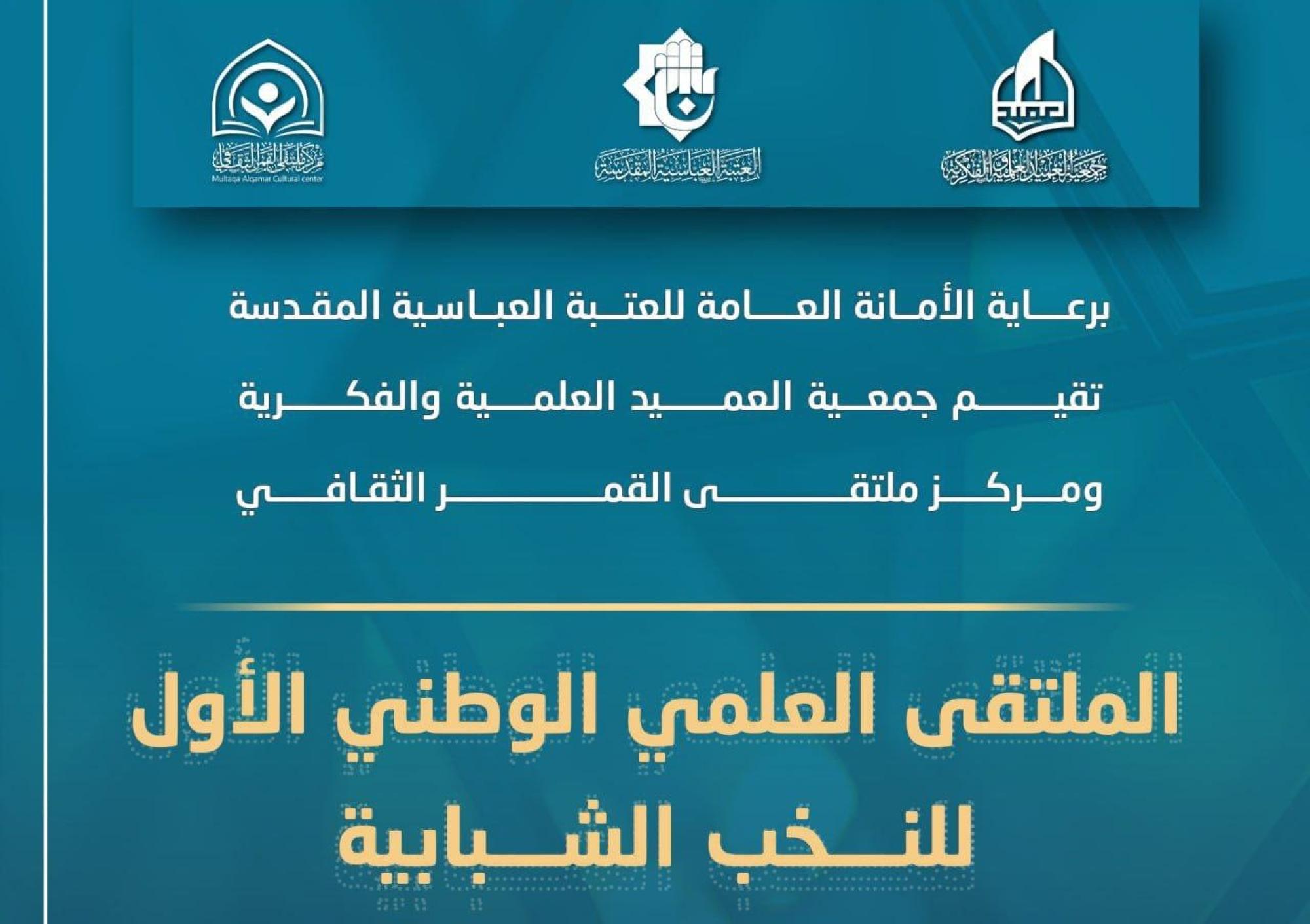
جمعيّة العميد وقسم الشؤون الفكريّة تدعوان الباحثين للمشاركة في الملتقى العلمي الوطني الأوّل
|
|
|
|
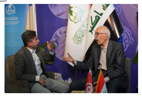
الأمين العام المساعد لجامعة الدول العربية السابق: جناح جمعية العميد في معرض تونس ثمين بإصداراته
|
|
|
|
المجمع العلمي يستأنف فعاليات محفل منابر النور في واسط
|
|
|
|
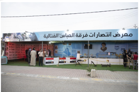
برعاية العتبة العباسيّة المقدّسة فرقة العبّاس (عليه السلام) تُقيم معرضًا يوثّق انتصاراتها في قرية البشير بمحافظة كركوك
|