آخر المواضيع المضافة

 النبات
الحيوان
الأحياء المجهرية
علم الأمراض
التقانة الإحيائية
التقنية الحيوية المكروبية
التقنية الحياتية النانوية
علم الأجنة
الأحياء الجزيئي
علم وظائف الأعضاء
الغدد
المضادات الحيوية
النبات
الحيوان
الأحياء المجهرية
علم الأمراض
التقانة الإحيائية
التقنية الحيوية المكروبية
التقنية الحياتية النانوية
علم الأجنة
الأحياء الجزيئي
علم وظائف الأعضاء
الغدد
المضادات الحيوية| Amyloid |


|
|
Read More
Date: 22-4-2021
Date: 25-4-2016
Date: 18-6-2021
|
Amyloid
Amyloid is an insoluble, proteinaceous, fibrous material associated with a number of prominent disease states (1), which can also form spontaneously in vitro from oligopeptides and denatured proteins. The disease states in which amyloid has been implicated as an important, or even causal, factor include Alzheimer's disease (see Amyloid Precursor Protein), the transmissible spongiform encephalopathies, non-insulin-dependent (type II) diabetes, and a number of polyneuropathies. It is thought that the extreme stability of amyloid fibrils permits their progressive accumulation in the extracellular spaces of vital organs whose functioning is thereby inhibited, leading to organ failure and death. The in vivo development of amyloid deposits from globular precursor proteins is linked to either genetic mutation, incorrect processing, or the abnormal accumulation of wild-type proteins. Probably the most remarkable feature of amyloid is that its molecular structure appears to be constant and independent of the protein precursors. For example, all amyloids, regardless of the disease involved or the source of the fibrils, share similar morphological (2), tinctorial (3), and structural (4, 5) properties. This implies that amyloid formation is not a simple aggregation process but a structural conversion of globular proteins to a state that can be incorporated into a particular type of fibrous protein structure. The amyloid diseases may therefore be defined as diseases of protein misfolding.
At least 15 different proteins can form amyloid fibrils in vivo (1), and recently the involvement of amyloid fibrils in Huntingdon's disease has also been demonstrated (6). In the electron microscope, amyloid fibers are about 100 Å (10 nm) in diameter and usually straight or only slightly curved. Closer examination shows the fibrils to be composed of a number of smaller diameter protofilaments, arranged in a more or less parallel array. In those fibrils so far examined, the protofilaments give the fibrils the appearance of hollow cylinders or ribbons when observed in cross section. Although the number, size, and arrangement of these constituent protofilaments appear to differ somewhat, the ribbons may be merely unrolled or unformed cylinders, and hence the variation may not be so great as appears. The molecular structure of the amyloid fibrils has been established by X-ray fiber diffraction. The first diffraction patterns (7, 8) showed the intense 4.7 Å (0.47 nm) meridional and 10 Å (1 nm) equatorial reflections that have been subsequently shown to characterize all amyloid X-ray patterns. These reflections indicate that the molecular structure of amyloid is composed of beta-sheets arranged parallel to the fiber axis, with their constituent beta-strands at right ngles to the axis of the fibril (4, 9). This so-called “cross-b” structure is quite different from the more common insect silk chorions, whose fibers are formed from b-sheets having their b-strands parallel to the fiber axis. The first use of intense synchrotron X-ray sources on amyloid (10) extended the observable X-ray pattern to 2 Å (0.2 nm) and revealed how this “classic” amyloid model could be reconciled with low-energy twisted b-sheets (11). These new data revealed a previously unobserved repeat distance of 115 Å (11.5 nm) along the fiber axis of the transthyretin amyloid of familial amyloidotic polyneuropathy (FAP), which was proposed (10) to correspond to the repeat distance of a complete helical turn of a twisted b-sheet, whose helix axis was parallel to the fiber axis. The basic helical unit therefore consists of a segment of b-sheet whose 24 b-strands complete one helical turn. This beta-helix model of the molecular structure of the amyloid protofilament is shown in Figure 1. Given that all amyloid fibrils share common morphological, tinctorial, and structural properties, it is reasonable to see the b-helix model as characteristic of all amyloid protofilaments. The b-helix model is distinct from other fibrous proteins, showing that the amyloid structure is unique. Its features enable low energy twisted b-sheets to be incorporated in a linear fibril in such a way that continuous b-type hydrogen bonding can be extended over the total length of the fibril. This, together with the opportunity for a continuous hydrophobic core to stabilize the b-sheet interactions along the length of the fibril, can be reasonably held to account for the known extreme stability of amyloid fibrils, which is central to their role in disease.
Figure 1. Drawing of one complete turn of the proposed b-helix structure of the amyloid fibril. The arrows represent b-strands and are shaded to represent the different b-sheets. The direction of the arrows has no significance, and no b-strand connections are shown.
A remarkable property of amyloid fibrils is that they appear to be very similar whatever their source. This has recently been critically tested (5) by showing that the high resolution synchrotron X-ray patterns from eight different amyloids, of both disease and synthetic origins, have the same structural characteristics. This observation suggests that amyloid is a highly stable generic structure capable of accommodating proteins and peptides with a wide range of chain lengths and structures within a common fibrillar form. The repetition of b-strands in the fibril model allows polypeptide chains of different lengths to occupy the same structural framework. For example, a 10-residue peptide could form a single b-strand of the amyloid structure, consistent with the known lower limit on the length of peptides capable of forming amyloid, while longer polypeptide chains could fold back and forward on themselves, thereby forming a number of consecutive b-strands of the amyloid b-sheets. The need for loops of chain of different lengths or conformation between successive b-strands to maximize the b-strand propensity, and to generate a stable hydrophobic core between the b-sheets, is likely to give each fibril a characteristic structure within the core amyloid framework. Thus it is probably realistic to consider amyloids as a closely related family of fibrils, each with its own detailed structural differences, while maintaining a similar overall core structure.
The nature of the processes that convert the globular, soluble protein precursors into amyloid fibrils, known as either amyloidogenesis or fibrillogenesis, are important, as they underpin the development of amyloid diseases and represent a novel field of scientific investigation. It is evident that some of the proteins forming the predominantly b-structured amyloid fibrils are themselves largely a-helical in their normal soluble states, for example, lysozyme, the prion protein, and the Ab peptide in Alzheimer's disease. The nature of this structural rearrangement has been particularly studied in two amyloidogenic proteins: transthyretin (TTR) and lysozyme. Transthyretin amyloidosis is associated with familial amyloidotic polyneuropathy (FAP) and senile systemic amyloidosis (SAA). Both the wild-type protein (SAA) and more than 50 different genetic variants (FAP) (13) give rise to amyloid. That so many variants of transthyretin are amyloidogenic (nearly 40% of all its residues), suggests that the structure of this protein allows a particularly facile transformation into the fibrillar form. As transthyretin is a 55-kDa homotetramer, with the four monomers associating in such a way as to generate a stack of four 8-stranded b-sheets (14), there is a superficial structural similarity with amyloid fibril (10). Nevertheless there is evidence that the tetrameric form of TTR is not a building block for the fibril (15), and the discovery of a molecular hotspot in the pattern of amyloidogenic variants (16) suggests that structural change to the monomer is also required prior to its incorporation into the fibril. It is therefore evident that even all-b proteins, such as TTR, must undergo a significant degree of structural rearrangement to form amyloid.
The human lysozyme molecule has about 35% a-helix and only 10% b-sheet structure, segregated into an a- and a b-domain. Hence the structural changes that must accompany its incorporation into the predominantly b-structured amyloid fibril will need to be extensive. Two genetic variants of human lysozyme are known to form amyloid in vivo, Ile56Thr and Asp67His (17) .A thorough biophysical study of these variants has highlighted major differences in their stability and folding behavior as compared with the wild type (18). It has generated a possible mechanism of the a- to b-structural transformation of fibril formation of lysozyme, shown in Figure 2 , which may be applicable mutatis mutandis to other amyloidogenic proteins. Thermal denaturation studies show that the variants unfold at temperatures at least 10°C lower than wild-type lysozyme and, unlike the wild type, do not regain activity when returned to physiological conditions. Fourier transform infrared spectroscopy of Asp67His lysozyme shows a significant gain in b-structure and loss of a-structure in the soluble material, demonstrating that an a-to-b structural interconversion is associated with fibril formation. Detection of an unfolding intermediate displaying the characteristic binding to the hydrophobic dye 1-anilinonaphthalene sulfonic acid (ANS) is consistent with a molten globule-like intermediate, as shown in Figure 2.
Hydrogen exchange shows that the flexibilities of the native folds of the variants have been dramatically increased by the mutations. Inspection of the X-ray crystallography structures of the two variants (18) suggests that the key to both amyloidogenic mutations in human lysozyme lies in the effect they produce at the interface between the a- and b-domains, with the result that domain adhesion may be weakened. Both variant lysozymes unfold dramatically faster than the wild-type protein, because the docking of the two domains, required for achieving the final rigid protein fold (19) is compromised by the presence of a threonine side-chain in the place of the wild-type hydrophobic anchoring residue, isoleucine.

Figure 2. Schematic drawing of the possible mechanism of fibril formation in human lysozyme. (—) b-sheet structure; (—) a-helical structure. A partly-folded, molten-globule form (II), distinct from the native (I), and denatured (III(forms, self-associates through the b-domain (IV) to initiate fibril formation. This provides a template for further deposition and the development of a b-sheet core structure of the amyloid fibril (V). Undefined structures are shown as broken lines.
Detailed study of the structure and behavior of variants of lysozyme has led to the proposal that transient populations of amyloidogenic proteins in a molten-globule state that lacks global cooperativity are an important feature of the conversion from a soluble to the fibrillar state (18). These observations lead to a model of the mechanism for amyloid formation for amyloidogenic lysozyme shown in Figure 2. Such a mechanism may also operate for other amyloidogenic proteins. For example, the structure of the 121–231 domain of the prion protein (20) also demonstrates that mutations associated with prion disease are involved in the maintenance of the hydrophobic core, which may be related to the earlier suggestion of a molten globule intermediate in the formation of aggregates in scrapie (21). Recent evidence supports the hypothesis that conformational plasticity is a key feature in prion fibril formation. A similar observation has been made for the Ab protein forming the amyloid in Alzheimer's disease (22). The mechanism for helix-to-sheet conversion in lysozyme, proceeding from soluble forms of the amyloidogenic precursor proteins through transient populations of intermediates with the characteristics of molten globules, and on to intermolecular b-sheet association, seems to parallel the processes in the aggregation of prions and the Ab amyloid peptides, and it may occur generally in the amyloidoses. Studies of these processes is certain to extend our knowledge of protein folding behavior into new areas and coincidentally address some of the more intractable diseases of our times.
References
1. M. B. Pepys (1994) in Santer''s Immunologic Diseases (M. M. Frank, K. F. Austen, H. N. Claman, and E. R. Unanue, eds.), Little, Brown & Co., Boston, pp. 637–655.
2. A. S. Cohen, T. Shirahama, and M. Skinner (1981) in Electron Microscopy of Protein, Vol. 3 (I. Harriss, ed.), Academic Press, London & New York, pp. 165–205.
3. G. G. Glenner, E. D. Eanes, and D. L. Page (1972) J. Histochem. Cytochem. 20, 821–826.
4. G. G. Glenner (1980) N. Eng. J. Med. 303, 1283–1292.
5. M. Sunde, L. C. Serpell, M. Bartlam, P. E. Fraser, M. B. Pepys, and C. C. F. Blake (1997) J. Mol. Biol. 273, 729–739.
6. E. Scherzinger et al. (1997) Cell 90, 549–558
7. E. D. Eanes and G. G. Glenner (1968) J. Histochem. Cytochem. 16, 673–677.
8. L. Bonar, A. S. Cohen, and M. Skinner (1967) Proc. Soc. Exp. Biol. Med. 131, 1373–1375.
9. J. H. Cooper (1976) in Amyoidosis (O. Wehelius and A. Paternak, eds.), Academic Press, London & New York, pp. 61–68.
10. C. C. F. Blake and L. C. Serpell (1996) Structure 4, 989–998.
11. C. Chothia (1973) J. Mol. Biol. 75, 295–302.
12. L. C. Serpell, M. Sunde, P. E. Fraser, P. Luther, E. Morris, E. Sandgren, E. Lundgren, and C. C. F. Blake (1995) J. Mol. Biol. 254, 113–118.
13. M. J. M. Saraiva (1995) Hum. Mutat. 5, 191–196.
14. C. C. F. Blake, M. J. Geisow, S. J. Oatley, B. Rerat, and C. Rerat (1978) J. Mol. Biol. 121, 339356- .
15. W. Colon and J. W. Kelly (1992) Biochemistry 31, 8654–8660.
16. L. C. Serpell, G. Goldsteins, I. Dacklin, E. Lundgren, and C. C. F. Blake (1996) Amyloid: Int. J. Exp. Clin. Invest. 3, 75–85.
17. M. B. Pepys et al. (1993) Nature 362, 553–557.
18. D. R. Booth et al. (1997) Nature 385, 187–793.
19. S. D. Hooke, S. E. Radford, and C. M. Dobson (1994) Biochemistry 33, 5867–5876.
20. R. Riek, S. Hornemann, G. Wider, M. Billeter, R. Glockshuber, and K. Wüthrich (1996) Nature382 , 180–182 .
21. J. Safar, P. Roller, D. Gajdusek, and C. Gibbs (1994) Biochemistry 33, 8375–8383
22. C. Soto, E. Castano, B. Frangione, and N. Inestrosa (1995) J. Biol. Chem. 270, 3063–3067.


|
|
|
|
للعاملين في الليل.. حيلة صحية تجنبكم خطر هذا النوع من العمل
|
|
|
|
|
|
|
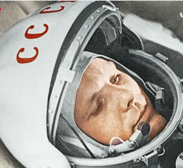
"ناسا" تحتفي برائد الفضاء السوفياتي يوري غاغارين
|
|
|
|
|
|
|
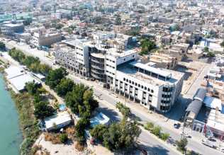
نحو شراكة وطنية متكاملة.. الأمين العام للعتبة الحسينية يبحث مع وكيل وزارة الخارجية آفاق التعاون المؤسسي
|
|
|