Aminopterin, Methotrexate, Trimethoprim, and Folic Acid
Aminopterin, methotrexate, and trimethoprim are all analogs of folic acid (Fig. 1) that antagonize folate-dependent metabolic pathways (1, 2). Folate is a water-soluble vitamin. Although some bacteria synthesize folate, mammalian cells cannot, and consequently it is an absolute dietary requirement. Reduced folates function as cofactors in many metabolic processes common to nearly all cells, such as thymidylate and purine synthesis and donation of methyl groups (Fig. 2) (1.( Folates gain entry into mammalian cells by either the reduced folate carrier (RFC) and/or a hydrophobic membrane-associated folate receptor (FR) found in placental, choroid plexus and kidney cells (3). The FR is unidirectional and, once inside, cellular retention of folate is enhanced by its polyglutamation, catalyzed by the enzyme folylpolyglutamyl synthetase (FPGS) (4). FPGS adds up to six or seven glutamic acid residues via an unusual peptide bond through their g-carboxyl group, rather than the normal a. The number of glutamic acid residues added may play a role in regulating and distributing reduced folates, and in guaranteeing their availability as cofactors. Polyglutamation also increases the affinity of folate for folate-dependent enzymes, such as thymidylate synthase (TS), aminoimidazole carboxamide ribonucleotide and glycinamide ribonucleotide transformylases. The latter two are involved in purine synthesis. In cells synthesizing DNA, 5,10-methylenetetrahydrofolate serves in thymidylate synthesis as a methyl group donor for converting dUMP to dTMP. This is the only reaction in which tetrahydrofolate is partially oxidized to dihydrofolate (1, 4). Dihydrofolate reductase (DHFR) is the critical enzyme involved in converting dihydrofolate back to tetrahydrofolate, thus maintaining reduced folate pools to serve as one-carbon group carriers (see Fig. 2).
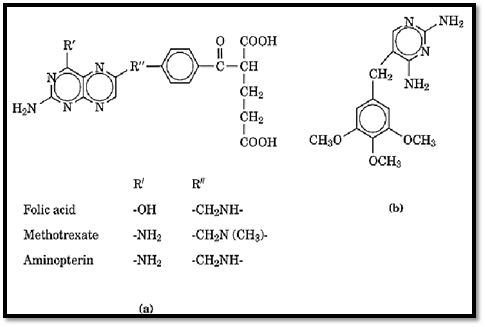
Figure 1. (a) The structures of folic acid and folate analogs: folic acid and analogs; (b) the structure of trimethoprim.
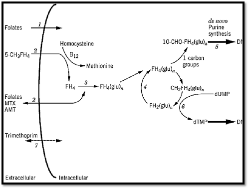
Figure 2. Folate transport, accumulation, and target enzymes. (1) folate receptor (FR); (2) reduced folate carrier (RFC), which is bidirectional; (3) folylpolyglutamyl synthetase (FPGS); (4) dihydrofolate reductase (DHFR); (5) aminoimidazol carboxamide ribonucleotide and glycinamide ribonucleotide tranformylases; (6) thymidylate synthase (TS); (7) passive diffusion of lipid-soluble compounds. FH4 and FH4 (glu)n, tetrahydrofolate with and without the added glutamyl residues FH2, dihydrofolate; CH2FH4, 5,10 methylene-FH4; 10-CHO-FH4, 10-formyl FH4; B12, vitamin B12 [Adapted from Hum and Kamen (1996) Investigational New Drugs 14, 110–111.]
Folate homeostasis is recognized as important. The structure of DHFR has been determined by X-ray crystallography. One proposed mechanism of resistance to methotrexate involves DHFR gene amplification (5-7). The FR is overexpressed in some carcinomas, and its gene has been localized to the 11q13 region (3). Human FPGS has been cloned and mapped to chromosome 9q (4). The complementary DNA for RFC has been cloned and the RFC gene localized to the long arm of chromosome 21 (8-11).
1. Methotrexate (MTX(
Folate analogs entered cancer therapy in the 1940s, when aminopterin was successfully used to induce temporary remissions in children with acute lymphoblastic leukemia (ALL) (12). Because of a better therapeutic index, MTX eventually emerged as the antifolate used clinically in treating cancers, such as leukemias, lymphomas, osteosarcoma, breast cancer, choriocarcinoma, head and neck cancers, and nonmalignant disorders, such as arthritis and asthma (4, 13).
Like folates, MTX enters mammalian cells via the RFC. MTX has an apparent dissociation constant in the micromolar range, and via the FR has higher affinity in the nanomolar range (3). Cellular retention of MTX is enhanced by polyglutamation, which also enhances the affinity of the drug for enzymes. Once inside the cell, MTX acts as a tight-binding competitive inhibitor of DHFR. This leads to an accumulation of dihydrofolate and depletion of the reduced folate pools in cells actively making dTMP via the de novo pathway. Accumulated dihydrofolate polyglutamates are also inhibitors of TS and the enzymes involved in the de novo synthesis of purines (1). The resulting imbalance of nucleotides causes base substitutions, which lead to errors in DNA synthesis and ultimately to cell death.
2. Aminopterin (AMT(
Aminopterin (AMT) was the first antifolate drug used clinically in childhood ALL (12) (see previous). Although more potent than MTX, in preclinical studies the toxicity of AMT was more severe and more unpredictable (13-15). Transport and metabolic studies in vitro have shown that AMT is the preferred substrate (16, 17). This results in greater AMT accumulation at lower concentrations and more complete polyglutamation, leading to improved cellular retention for the cytotoxic effect.
The greater potency of AMT compared to MTX has led to renewed clinical interest. AMT may find a role in treating patients with resistant or refractory malignancies or those patients in whom in vitro studies indicate AMT is the better choice on the basis of metabolism and accumulation (16).
3. Trimethoprim
Trimethoprim is an antibacterial agent developed in the 1950s. Studies by Hitchings (2) showed that its mechanism of action is the competitive inhibition of DHFR. Unique to trimethoprim is its much greater affinity (50,000- to 100,000-fold) for bacterial DHFR than for mammalian DHFR (18, 19).
Trimethoprim is lipid-soluble and enters cells rapidly without requiring specific transport mechanisms. Its selective toxicity is further enhanced by the ability of folinic acid to reverse even the slight effects of trimethoprim on mammalian cells, whereas bacterial cells, unable to transport folinic acid, are not rescued by folinic acid administration (19, 20). In addition to its antibacterial activity, the drug has activity against Pneumocystis carinii, an opportunistic infection of the lungs encountered in severely immunocompromised patients (especially HIV patients).
References
1. M. C. Hum and B. A. Kamen (1996) Investigational New Drugs 14, 110–111.
2. G. H. Hitchings (1973) J. Infect. Dis. 128(suppl), S433–S436.
3. S. Weitman, R. G. W. Anderson, and B. A. Kamen (1994) In Vitamin Receptors: Vitamins as Ligands in Cell Communities (K. Dakshinamurti, ed.), Cambridge University Press, Cambridge, UK, pp. 106–136.
4. T. A. Garrow, A. Admon, and B. Shane (1992) Proc. Natl. Acad. Sci. USA 89, 9151–9155.
5. F. W. Alt et al. (1978) J. Biol. Chem. 253, 1357–1370.
6. J. R. Bertino (1993) Ode to Methotrexate, J. Clin. Oncology 11(1), 5–14.
7. E. Chu and C. H. Takimota (1993) In Principles & Practice of Oncology (V. T. De Vita, S. Hellman, and S. A. Rosenburg, eds.), Lippincott, Philadelphia, pp. 358–374.
8. J. A. Moscow et al. (1995) Cancer Res. 55, 3790–3794.
9. P. D. Prasad et al. (1995) Biochem. Biophys. Res. Commun. 206, 681–687.
10. S. C. Wong et al. (1995) J. Biol. Chem. 270, 17468–17475.
11. T. L. Yang-Feng et al. (1995) Biochem. Biophys. Res. Commun. 210(3), 874–879.
12. S. Farber et al. (1948) N. Engl. J. Med. 238, 787–793.
13. A. Goldin et al. (1955) J. Natl. Cancer Inst. 15, 1657–1664.
14. F. M. Sirotnak and R. C. Donsbach (1975) Biochem. Pharmacol. 24, 156–158.
15. F. S. Philips et al. (1973) Cancer Res. 33, 153–158.
16. A. Smith et al. (1996) Clin. Cancer Res. 2(1), 69–73.
17. B. G. Rumberger, J. R. Barrueco, and F. M. Sirotnak (1990) Cancer Res. 50, 4639–4643.


