آخر المواضيع المضافة

 النبات
الحيوان
الأحياء المجهرية
علم الأمراض
التقانة الإحيائية
التقنية الحيوية المكروبية
التقنية الحياتية النانوية
علم الأجنة
الأحياء الجزيئي
علم وظائف الأعضاء
الغدد
المضادات الحيوية
النبات
الحيوان
الأحياء المجهرية
علم الأمراض
التقانة الإحيائية
التقنية الحيوية المكروبية
التقنية الحياتية النانوية
علم الأجنة
الأحياء الجزيئي
علم وظائف الأعضاء
الغدد
المضادات الحيوية| The Cell and Cell Cycle |


|
|
Read More
Date: 11-11-2015
Date: 27-10-2015
Date: 11-11-2015
|
The Cell and Cell Cycle
INTRODUCTION
The cell, the simplest living structure capable of independent existence, was first identified in 1663 by an English scientist Robert Hooke. It was not until 1838, that Schleiden and Schwann announced that the cell was the basic structural unit and functioned according to definite laws. The forms and functions of cells are diverse. They are controlled by genes, which lie on the chromosomes present in the cell nucleus. The chromosomes are involved in cell division as well as reproduction. To understand the basis of various genetic disorders, the study of cell structure and cell cycle is necessary. In unicellular organisms, a single cell carries out all the functions necessary for its survival. In higher organisms, however, cells associate to form colonies where different cells are allocated various functions, these being interdependent. The aggregates of cells, which have specialized functions, form different tissues, like blood, nervous tissue, bone and muscles. These tissues combine to form specialized organs such as the kidneys, heart and lungs. These in turn are grouped into functioning systems, like the urogenital, cardiovascular and respiratory systems.
COMPONENTS OF A CELL
Each cell has three basic components, (the cytoplasm, a cell membrane, which forms the cell wall, and a centrally placed body, the nucleus (Fig. 1).

Fig 1: Components of a cell
Cytoplasm
The cytoplasm is a colloidal matrix composed of water and inorganic and organic compounds. Amongst the inorganic molecules are sodium, potassium, calcium, magnesium, bicarbonate and phosphates in trace amounts.
Organic molecules that impart colloidal property to the cytoplasm are monomers such as nucleotides, amino acids, monosaccharides and fatty acids along with their polymers, nucleic acids, proteins, polysaccharides and lipids. These constitute the macromolecules making up the major structural and functional units of the cell. The functions of each unit are different. Some proteins give structural support, like actin and myosin of the muscle and keratin of hair and nails. Some are involved in catalysis of metabolic reactions. Complex cellular reactions involve hormones, receptors and growth factors.
Nucleic acids are the repositories of genetic information and act as templates for the synthesis of proteins. Nucleic acids are of two types, deoxyribonucleic dcid (DNA) and ribonucleic acid (RNA). Purines and pyrimidines, are composed of a five- carbon sugar (pentose), a phosphate group, and a cyclic nitrogen compound. Purines are adenosine and guanine and pyrimidines are cytosine and thymine. Thymine is replaced by uracil in RNA. The sugar moiety in DNA is deoxyribose and that in RNA is ribose.
Lipids encompass a diverse group of compounds that are soluble in organic solvents. These include phospholipids in the cell membrane, sphingolipids in the nervous tissue, glycolipids in myelin sheath and steroids including male and female hormones, bile and adrenocortical hormones.
Cell Membrane
The cell membrane, also termed plasmalemma, defines the cellular boundary and acts as a physical barrier for cellular contents. It consists primarily of phospholipids and proteins. The membrane has selective permeability, which allows them to and fro passage of molecules. This is achieved by three mechanisms: passive diffusion, active transport and enclosure.
Passive diffusion is a term used to describe movement of substances from a region of high concentration to regions of lower concentration. Active transport requires energy and moves substances against a concentration gradient. Enclosure in vesicles that move substances into the cells is called endocytosis or pinocytosis, and out of cells is called exocytosis. Water moves freely across the membrane in both directions.
Glycoproteins are present on the protein lipid membrane surface. Their function is cell adhesion. Glycoproteins also have antigenic properties, and in red cells they determine blood groups.
Light microscopy has limitations in further identification of structures, which can be observed only by electron microscopy (EM). Some of these structures include the smooth endoplasmic reticulum, which functions in lipid metabolism. Rough ER, which has ribosomes attached to it, are the site of protein synthesis. Golgi apparatus is involved in the modifying, sorting and packing of molecules for secretion or delivery to other organelles. Lysosomes are vesicles containing digestive enzymes involved in the disposal of native or foreign waste products. Mitochondria are the powerhouses of cells, where oxidation of nutrients occurs to provide energy for synthesizing ATP. Structurally, mitochondria are small bodies with a double membrane. The inner membrane is folded into numerous projections called cristae, where oxidation of nutrients takes place. The other bodies in the cytoplasm are centrioles or basal bodies. Centrioles are responsible for the formation of spindle fibres, which separate chromosomes to respective daughter cells during cell division, and aid in the formation of cilia and flagella, which are needed for cell motility.
Nucleus
The nucleus carries the hereditary material, DNA, which determines specific functions and characteristics of a cell. The DNA lies in condensed form in linear arrays called chromosomes. Organisms with cells having a nucleus are called eukaryotes, and they are plants, animals and humans. Those without a proper nucleus are called prokaryotes, for example, bacteria. In prokaryotes the genetic material lies in the cytoplasm.
Cells lacking nuclei have limitations in their metabolic activity. When the cell goes through cell cycle, its appearance differs. The metaphase stage cell has its nucleus in a condensed spherical body and is darkly stained (heterochromatin). In interphase, two types of chromatin are seen.
The nucleus has an outer nuclear membrane and contains nucleoli and chromatin. The nuclear membrane or envelope is a double membrane with ribosomes attached to the outside. The membrane at many sites is continuous with the ER. When a cell divides, the nuclear membrane disappears. Within the nucleus, there is nucleolus and chromatin. The size and number of nucleoli vary with the cell type and the metabolic state of the cell. The nucleoli are larger in rapidly dividing cells and in cells with active protein synthesis. All the ribosomes in the cytoplasm originate in the nucleolus. Each nucleolus is formed along the nucleolar-organizing region of one or more specific chromosomes and is recognizable during cell division. The nucleolus is composed of RNA, protein and some amount of DNA.
The chromatin is composed of DNA, proteins (mainly histones), RNA and polysaccharides.
Euchromatin and heterochromatin
During the cell cycle, chromosomes show a property of condensation (coiling) and decondensation. Maximum condensation occurs at metaphase. The staining intensity of the chromosomes varies owing to this property of condensation called heteropyknosis. More heavily stained parts of the chromosome are called areas of positive heteropyknosis and light areas are those of negative heteropyknosis. The chromatin in these variable regions is called heterochromatin and in the other regions in the cell it is called euchromatin. Heterochromatin is of two types, facultative and constitutive. The inactive X chromosome in the female gets condensed and is facultative heterochromatin while the other differentially staining areas of the chromosome seen in banding are constitutive heterochromatin.
X CHROMATIN AND Y CHROMATIN X Chromatin
In 1949 Barr and Bertram in their experiments on cat nerve cells, observed a peculiar body, which they called as paranucleus (now called the Barr body), and this was present only in female cats. In 1961, Mary Lyon put forth a hypothesis that one of the X-chromosomes of females is inactivated and this chromosome could be of maternal or paternal origin. The inactivation is stable and occurs at embryogenesis. It was hypothesized that this was to compensate for the extra gene products produced in females who have two X-chromosomes and is called dosage compensation.
As a result of this random inactivation of X-chromosomes, females are always mosaic for the genes located on the X chromosome. The inactivated X is observed as a darkly stained body in the nucleus attached to the nuclear membrane (Fig. 2A). It is either triangular, oval or dumbbell shaped and is always one per each inactivated X chromosome. Males with XXY complement will show presence of one Barr body or females with XXX syndrome will have two Barr bodies. This test along with Y chromatin studies can be offered as a provisional diagnostic test in ambiguous genitalia. The inactivation centre is believed to reside on the Xq13 region on the long arm of the X chromosome.
Y Chromatin
In a normal male, the sex chromosomal pattern is XY The Y chromosome belongs to the G group of chromosomes and is easily distinguishable from chromosome 21 and 22. The Y chromosome does not have a satellite and the long arms are straight. They do not diverge like long arms of chromosomes 21 and 22). The length of this segment varies. The Y chromosome is transmitted from father to son and the length of the Y can be studied as a family marker. When the buccal smears, peripheral blood smear or smears from seminal fluid are stained with a quinacrine dye, this fluorescent segment can be visualized in the interphase nuclei as a brightly fluorescent body called as Y chromatin (Fig. 2B).
The role of satellite DNA is becoming increasingly important in techniques like fluorescent in situ hybridization (FISH). Repetitive DNA found in constitutive heterochromatin is called satellite DNA. Satellite DNA has highly repetitive sequences. A substantial portion of each fraction is made up of a single family of simple repeats. There are variations from mutations, sharing one to a few base pair differences. The alpha and beta satellite DNA is found at the centromere of all chromosomes. Satellite probes identify the centromeric regions of specific chromosomes and are used to identify aneuploidies or X and Y chromosomes in uncultured cells.

Figs 2A and B: Sex Chromatin in buccal mucosa. (A) X chromatin (B) Y chromatin
The cell cycle
For growth, cells need to multiply. In this process the cell mass increases, duplication of the genetic material occurs, and then cell division takes place. This assures that each newly formed daughter cell receives an equal component of genetic material. These orderly mannered stages of cell division are referred to as the cell cycle.
The cell cycle is divided into four phases (Fig. 3):
Mphase: This is a relatively brief phase in which mitosis and cell division occurs.
G1 phase: G1 phase follows mitosis. This is the gap phase, which covers the longest part of the cell cycle.
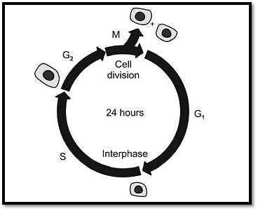
Fig. 3: Stages in cell cycle
S phase: This is the synthesis phase, which, in contrast to prokaryotes, is the only phase in which DNA is synthesized in eukaryotes.
G2 phase: The cell, which has become tetraploid, now prepares itself for division. Two processes are involved in a cell division the first is called mitosis, where nuclear division occurs and the second cytokinesis, where changes occur in the cytoplasm, including division of the cell proper. G2 is a relatively short phase. Once the cell enters the M phase, again a new round of cell division begins
Typically, cells in culture complete a cell cycle within 1624 hr. This may vary from 8 hr to upto to 100 days or mors for different types of cells. This variation usually occurs in the Gx phase. Cells that have differentiated terminally never divide; they enter the Go phase also known as the quiescent phase. For a cell with a 24-hour cycle, Gx phase requires 10 hours, S phase requires 9 hours, G2 requires 4 hours and mitosis 1 hour. A cell’s irreversible decision to proliferate is made during the Gx phase. Cells remain quiescent if nutrients are inadequate or if they are in contact with each other (contact inhibition).
DNA synthesis may be induced by (i) various agents such as carcinogens or tumour viruses, which trigger uncontrolled cell proliferation (as seen in cancer) (ii) Surgical removal of a tissue which results in rapid regeneration (iii) mitogens which are proteins that bind to cell surface receptors and induce cell division (iv) certain cytoplasmic factors present in growing cells which stimulate DNA synthesis.
Mitosis
Mitosis is a continuous process, and is subdivided into 4 stages, prophase, metaphase, anaphase, and telophase. Between cell division, cells are said to be in interphase (Fig. 4). The type of tissue, temperature and nutritional health of cell determine the relative length of each stage.
Interphase: In late interphase, cells prepare to undergo mitosis. The nucleus assumes a reticulate appearance due to the maximally extended, uncoiled chromosomes. There is often a single nucleolus at this stage. A centrosome encompassed by astral rays and containing a medium centriole is seen at the surface of the nuclear envelope.
Prophase: Until prophase begins, it is usually not apparent that a cell is about to divide. Generally, the cell enlarges relative to the neighbouring cells.
Early prophase: During the early part of prophase divided chromosomes separate and take their positions at opposite poles. The chromosomes now coil into compact structures and appear shorter and thicker. The nucleoli disperse.
Late prophase: At the end of prophase, chromosomes become clearly visible and nucleoli disappear.
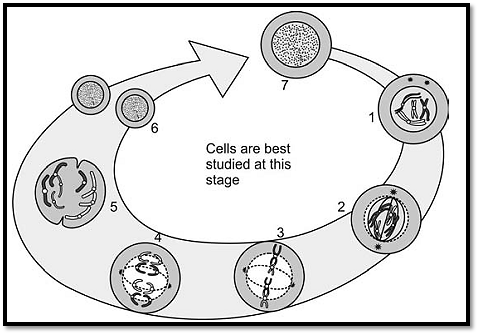
Fig. 4 : Normal cell division stages in mitosis 1. Prophase 2. Prometaphase 3. Metaphase 4. Anaphase 5. Telophase 6. Interphase
Prometaphase: This is the portion of prophase immediately preceding metaphase. The chromosomes attain their maximum thickness and minimum length. Each chromosome that has split longitudinally for most of its length remains connected at a single point at the centromere. These separated chromosomes are called sister chromatids. The nuclear membrane begins to break down and chromosomes are left in the cytoplasm. A mitotic apparatus begins to assemble, and chromosomes start taking their positions at the equatorial plane after attachment of the centromeres of each chromosome to spindle fibres. The spindle apparatus seen now, consists of centromeres, their encompassing astral rays, a gelatinous spindle made up of fibres extending between centrosomes and traction fibres extending from each centrosome to the chromosomal centromere.
Metaphase: This is usually a very short stage. Chromosomes can be seen aligned equatorially in the mitotic apparatus and can be best studied and counted at this time.
Anaphase: During this phase the separation of chromosomes begins.
Early Anaphase: Each centromere divides longitudinally, thus converting two chromatids of the chromosomes into two daughter chromosomes. These daughter chromosomes disjoin and gradually move to opposite poles. This occurs due to pulling of the chromosomes by traction, in a process called karyokinesis. The longer chromosomes may still be adhered at their distal ends.
Late Anaphase: Chromosomes are pulled towards the pole and as they move away from the centre and the cell membrane starts invaginating. This process is called cytokinesis.
Telophase: This phase begins when sister chromatids reach the poles. The cell membrane invaginates from the area opposite the spindle equator. This process, which begins in late anaphase ends here. The nuclear membrane is formed around the chromosomes thus separating them from the centriole and the rest of the cytoplasm. Chromosomes become uncoiled again and spindle fibres and astral bodies disappear. The centriole divides as the centrosome prepares for the next mitosis.
The sequential and purposeful actions of mitosis focus on the movements of the chromosomes to ensure that they are distributed equally. It is essential that each chromosome of the parent cell have an identical counterpart in each of the daughter cells.
Meiosis
Union of two haploid germ cells or gametes, an egg from the mother and a sperm from the father form the diploid zygote.
These haploid cells cannot form by mitosis, as a reduction in the number of parental chromosomes to half is required. This occurs by a process termed meiosis involving two divisions. Reduction is affected because the two divisions involve only a single replication. There is orderly distribution of these replications in meiosis. In most organisms, meiotic cells are segregated in specialized organs generally termed gonads. (i) The female cells (containing abundant stored food to nourish the embryo in its early stages) are termed eggs or ova. This type of meiosis is called oogenesis and takes place in the ovary, (ii) In male, these are called spermatozoa and are produced by spermatogenesis in the testes.
The history of male and female gametes is different but the sequence is same. In males and females, there are two successive meiotic divisions. Meiosis I is known as reduction division since the chromosome number is reduced to haploid by pairing of homologous chromosomes in prophase and their segregation at anaphase in this division (Fig. 5A). The X and Y pair only at the tip of their short arms, as that is the homologous region.
Meiosis I
Prophase I: This is a complicated process, and differs from the mitotic prophase in a number of ways with important genetic consequences. It is long and critical, and is usually studied as five different stages, throughout which the chromosomes continually condense and become shorter and thicker. The stages of prophase I are described below.
Leptotene: Leptotene is characterized by the first appearance of 46 chromosomes. The chromosomes, which have already replicated during the S phase, become visible as thin threads that begin to condense. The sister chromatids are so closely aligned, that they cannot be distinguished as separate. Unlike mitotic chromosomes, meiotic chromosomes have alternating thicker and thinner regions. The pattern of thick regions (chromosomes) is characteristic for each chromosome.
Zygotene: In this stage, the chromosomes start pairing along their entire length. This pairing is also called synapsis and is very precise. Electron microscopy reveals the synaptonemal complex to be a ribbon like tripartite structure containing protein. This complex is essential for crossing over, which is the exchange of homologous segments between non-sister chromatids of a pair of homologous chromosomes. Crossing over, which occurs in the subsequent step, is biologically and clinically significant.
Pachytene: In this phase, the chromosomes become much more tightly coiled and mono pronounced. Synapsis is complete and structures called tetrads (as they contain four chromatids) are seen. Crossing over takes place at this stage.
Diplotene: The homologous chromosomes in each bivalent structure begin to repel each other. Here, their centromeres remain attached to each other and the chromosomes are held together only at points where the crossover takes place. These sites are termed chiasmata.
Diakinesis: When the prophase is nearing the end, the chromosomes move onto the spindle, and the tetrads become very contracted and densely stained. Terminalization is completed here.
Metaphase I: As in mitosis, the nuclear membrane disappears, and a spindle forms. The chromosomes align themselves on the equatorial plane. Their centromeres are oriented towards different poles.
Anaphase I: It is characterized by the separation of the chromosomes that had formerly formed the bivalents.
One of each pair moves to one pole of the spindle and the other member to the other pole. This is termed disjunction. This results in sorting of maternal and paternal chromosomes in random combinations. The possible number of combinations is 223. The process of crossing over imparts more variety. Anaphase I is the most error-prone step in meiosis.
Telophase I: The centromeres remain intact. Hence the 23 chromosomes at each pole remain double stranded and are called dyads. A nuclear membrane is formed around each group of 23 dyads.
Cytokinesis: The cell divides into two haploid daughter cells and enters interphase. Cytokinesis differs in spermatogenesis and oogenesis. In spermatogenesis, the cytoplasm is almost equally divided between two spermatocytes, but in oogenesis, one product (the secondary oocyte) receives almost all the cytoplasm, and the other becomes the first polar body. Hers interphase is brief and there is no phase between the first and second meiotic divisions. After this phase, the chromosomes decondense again and meiosis II begins.
Meiosis ll
This is the second meiotic division. This is similar to mitosis except that the chromosome number of the cell entering this phase is haploid (Fig. 5B). On completion of this division, four haploid cells, each containing 23 chromosomes is formed. Due to crossing over in meiosis I, the chromosomes of the daughter cells are not identical to those of the parent cell. Segregation of paternal and maternal forms of each gene takes place during either first or the second mitotic division, depending on whether they have been involved in a crossover event in meiosis I.
Chromosomal errors occur due to failure in the normal mitotic and meiotic divisions.
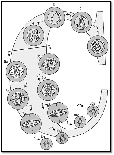
Fig. 5A: Stages of meiosis I. 1 through 4, stages of prophase I, 5a and b, metaphase I, 6a and 6b, anaphase I, 7a and 7b, telophase I and 8a1, 8a2, 8b1 and 8b2 represent the possible outcomes.
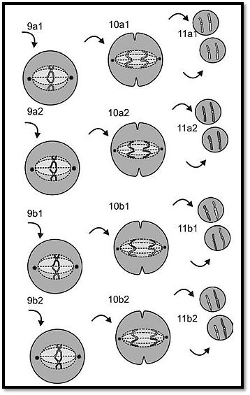
Fig. 5B: Stages of meiosis II, 9a1, 9a2, 9b1 and 9b2, anaphase II,10a1,10a2,10b1 and 10b2, stages of telophase II 11a1, 11a2,11b1,11b2 represent the possible outcomes.
Gametogenesis
Male and female gametogenesis have a basic difference in the process, and various errors can occur in the genetic material leading to genetic variations or defects in the offspring (Fig. 6).
Oogenesis
Primordial germ cells give rise to oogonia by 20-30 mitotic divisions. This process occurs in the first few months of embryonic life. At the end of three months of embryogenesis, the oogonia mature into primary oocytes and meiosis starts. At birth these primary oocytes enter a phase of maturation arrest, dictyotene and the ovum is suspended in the prophase stage till meiosis I, which is completed at the time of ovulation. A single secondary oocyte is then formed, and the other cell is called polar body. The secondary oocyte receives most of the cytoplasm. The process of meiosis II commences during fertilization. Oogonia are present in embryonic life and at each menstrual cycle one egg matures and is released. In the reproductive life of a female, from first the onset of menstruation to menopause, approximately 300 ova are released. The others become atretic. The fact that many ova are available for maturation in every cycle is taken advantage of in assisted reproductive technology, where per cycle with hormonal induction about 20 to 25 mature ova can be made available for aspiration. As the process of oogenesis is a lengthy procedure, advanced maternal age plays a great role in chromosomal aneuploidy (numerical defects). There is always a chance that during this period a primary oocyte is exposed to intrinsic or extrinsic factors, which can damage spindle formation and the repair process, resulting in non-disjunction.
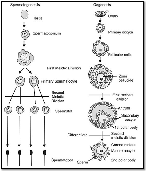
Fig. 6 : Normal gametogenesis A. Spermatogenesis B. Oogenesis
References
Purandarey , H. (2009) . Essentials of Human Genetics. Second Edition. Jaypee Brothers Medical Publishers (P) Ltd.


|
|
|
|
طبيبة تبدد 5 خرافات رئيسية عن تغذية الأطفال
|
|
|
|
|
|
|
أول شريحة مزروعة في دماغ إنسان تواجه مشكلة.. ماذا حدث؟
|
|
|
|
|
|
الأمين العام للعتبة الحسينية: ينبغي أن تحاط اللغة العربية بالجلالة والقدسية فهي سلاح الأمة وسبيل وحدتها ونهضتها
|
|
|
|
بالفيديو: الامين العام للعتبة الحسينية: مشروع الكابل الضوئي هو مشروع تنموي كبير سيرفع من سقف التنمية في محافظة كربلاء
|
|
|
|
بالفيديو: بحضور ممثل المرجعية العليا والامين العام للعتبة الحسينية.. جامعة الزهراء (ع) للبنات تحتفي بتخرج (دفعة طوفان الاقصى)
|
|
|
|
بالتعاون مع جامعة ليفربول وتستهدف مليون فحص مجاني... العتبة الحسينية تعلن عن موعد إطلاق حملة للكشف المبكر عن الأمراض السرطانية
|