آخر المواضيع المضافة

 النبات
الحيوان
الأحياء المجهرية
علم الأمراض
التقانة الإحيائية
التقنية الحيوية المكروبية
التقنية الحياتية النانوية
علم الأجنة
الأحياء الجزيئي
علم وظائف الأعضاء
الغدد
المضادات الحيوية
النبات
الحيوان
الأحياء المجهرية
علم الأمراض
التقانة الإحيائية
التقنية الحيوية المكروبية
التقنية الحياتية النانوية
علم الأجنة
الأحياء الجزيئي
علم وظائف الأعضاء
الغدد
المضادات الحيوية| Urinary system |


|
|
Read More
Date: 9-7-2021
Date: 1-8-2021
Date: 14-7-2021
|
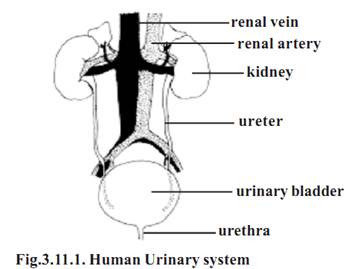
Urinary system
It is customary to link the organs of urinary excretion and reproduction as urinogenital system. The suitability of this concept is questionable.
The urinary and reproductive organs differ in their embryological origin and development. In postnatal human beings, the association between the components of the urinary and the reproductive systems is very much limited. Hence the urinary and reproductive systems are considered separately.
The urinary organs comprise, two kidneys (renes), ureters, the urinary bladder (vesica urinaria) and the urethra.
The kidneys
The kidneys are bean shaped organs. In fresh state the kidneys are reddish brown in colour. They lie on the posterior abdominal wall. In the abdomen, the right kidney is slightly lower than the left. It is because of the presence of liver superior to it. The kidneys are surrounded by adipose tissue. Each kidney is about 11 cm in length, 6cm in breadth and 3cm in anteroposterior dimensions. In adult males the average weight of kidney is about 150g (in adult female 135g).
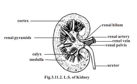
The inner margin of each kidney has a small depression called the hilum. The renal artery and nerves enter and the renal vein and the ureter exit at this region. The hilum opens into a cavity called the renal sinus.
Each kidney is enclosed by a fibrous connective tissue layer, called the renal capsule. Internally the kidney is divided into an outer cortex and an inner medulla. The medulla consists of several cone-shaped renal pyramids. Extensions of the pyramids called the medullary rays, project from the pyramids into the cortex. Extension of the cortex called renal columns, project between the pyramids. The tips of the pyramids are called the renal papillae. They are pointed toward the renal sinus. The renal papillae are surrounded by funnel shaped structures called the minor calyces. The minor calyces of several pyramids join together to form larger funnels called major calyces. There are 8-20 minor calyces and 2 or 3 major calyces per kidney. The major calyces converge to form an enlarged channel called the renal pelvis. The renal pelvis then narrows to form the ureter. The ureter leaves the kidney and gets connected to the urinary bladder.
Nephron.
The basic functional unit of each kidney is the nephron. There are approximately 1.3 million nephrons in each kidney. At least 450,000 of them must remain functional to ensure survival. Each nephron consists of an enlarged terminal end called the renal corpuscle, a proximal tubule, a loop of Henle and a distal tubule. The distal tubule opens into a collecting duct. The renal corpuscle, proximal tubule and distal tubules are in the renal cortex. The collecting tubules and parts of the loops of Henle enter the renal medulla.
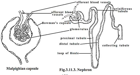
Most nephrons measure 50-55 mm in length. 15% of the nephrons are larger and they remain near the medulla. These are called the juxtamedullary nephrons. They have larger loops of Henle.
The renal corpuscle of the nephron consists of a Bowman’s capsule and a bunch of capillaries called the glomerulus.
In the Bowman’s capsule the outer and inner layers are called parietal and visceral layers respectively. The outer parietal layer is composed of simple squamous epithelium. The inner visceral layer surrounds the glomerulus. It consists of specialized cells called podocytes. The walls of the glomerular capillaries are lined with endothelial cells. There is a basement membrane between the endothelial cells of the glomerular capillaries and the podocytes of Bowman’s capsule. The capillary endothelium, the basement membrane and the podocytes of Bowman’s capsule make up the filtration membrane.
The glomerulus is supplied with blood by an afferent arteriole. It is drained by an efferent arteriole.
The cavity of Bowman’s capsule opens into the proximal tubule. The proximal tubule is also called the proximal convoluted tubule. It is approximately 14mm long and 60 µm in diameter.
Posteriorly the proximal tubule continues as the loop of Henle. Each loop has a descending limb and an ascending limb. The first part of the descending limb is similar in structure to the proximal tubule. The loops of Henle that extend into the medulla become very thin near the end of the loop. The first part of the ascending limb is also very thin and it consists of simple squamous epithelium, but it soon becomes thick. The distal tubules, also called the distal convoluted tubules are not as long as the proximal tubules.
Ureters and Urinary bladder
The ureters extend inferiorly from the renal pelvis. They arise medially at the renal hilum to reach the urinary bladder. The bladder is meant for temporarily storing the urine. The urinary bladder is a hollow muscular bag. It lies in the pelvic cavity. The size of the bladder depends on the presence or absence of urine. The bladder capacity varies from 120-320ml. Filling up to 500 ml is tolerated. Micturition will occur at 280ml. The ureters enter the bladder inferiorly on its posterolateral surface. The urethra exits the bladder inferiorly and anteriorly. At the junction of the urethra with the urinary bladder smooth muscles of the bladder form the internal urinary sphincter. Around the urethra there is another external urinary sphincter. The sphincters control the flow of urine through the urethra.
In the male the urethra extends to the end of the penis where it opens to the outside. In male the urethra is 18-20cm long. In the female the urethra is shorter. It is about 4 cm long and 6 mm in diameter.
References
T. Sargunam Stephen, Biology (Zoology). First Edition – 2005, Government of Tamilnadu.


|
|
|
|
دراسة: عدم ترتيب الغرفة قد يدل على مشاكل نفسية
|
|
|
|
|
|
|
علماء: تغير المناخ تسبب في ارتفاع الحرارة خلال موسم الحج
|
|
|
|
|
|
|
شعبة فاطمة بنت أسد للدراسات القرآنية تختتم دورة ينابيع الرحمة
|
|
|