آخر المواضيع المضافة

 النبات
الحيوان
الأحياء المجهرية
علم الأمراض
التقانة الإحيائية
التقنية الحيوية المكروبية
التقنية الحياتية النانوية
علم الأجنة
الأحياء الجزيئي
علم وظائف الأعضاء
الغدد
المضادات الحيوية
النبات
الحيوان
الأحياء المجهرية
علم الأمراض
التقانة الإحيائية
التقنية الحيوية المكروبية
التقنية الحياتية النانوية
علم الأجنة
الأحياء الجزيئي
علم وظائف الأعضاء
الغدد
المضادات الحيوية| The Circulatory System |


|
|
Read More
Date: 16-7-2021
Date: 12-7-2021
Date: 30-7-2021
|
The Circulatory System
The multicellular organization in animal world has resulted in the origin and evolution of circulatory system in animals. This arrangement facilitates internal transport of various substances to all organs and organ systems. Among majority of multicellular animals this system remains as a closed type. It has blood running inside closed blood vessels, the blood being pumped by heart. In man, as in all mammals there is a double circulation of blood. The primary circulation through pumping action of heart, supplies blood to all regions of the body. The blood later returns to the heart. It is called the systemic circulation or body circulation. A similar circulation carries blood to lungs for oxygenation and returns it back to the heart. It is called the pulmonary circulation.
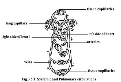
Systemic and Pulmonary circulations
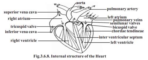
The most important component of this system is the heart. It is a large, muscular, valve structure having four chambers. The chambers are the right atrium, left atrium, right ventricle and left ventricle. Each atrium opens into a corresponding ventricle. The right and left chambers are separated by septa.
Systemic circulation: - The left atrium receives oxygenated blood from the lungs, through the pulmonary vein. When the atria contract, blood from the left atrium is forced into the left ventricle. Later by a contraction of the ventricle, the blood leaves the heart through the aorta. The aorta is the single systemic artery emerging from the heart. By successive branching, the aorta
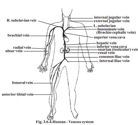
gives rise to hundreds of arteries taking blood to all regions of the body. As the branching happen, the arteries divide into numerous (4 x 10⁶) arterioles. In the target organs they produce four times as many capillaries. A similar number of venules converge into each other forming veins of increasingly larger size. Finally, only two veins, the superior and inferior vena cavae return the blood to the right atrium. Thus the course of blood from left ventricles through the body organs and back to the atrium forms the systemic circulation.
Pulmonary circulation: - The venous blood from right atrium is conducted to the right ventricle. The ventricle expels the blood via the pulmonary trunk to the lungs. The oxygenated blood later returns by the pulmonary veins to the left atrium. This circulation from right ventricle to the left atrium via the lungs is termed the pulmonary circulation.
Portal circulation:-In the systemic circulation the venous blood passing through spleen, pancreas, stomach and intestine is not carried back directly to the heart. It passes through the hepatic portal vein to the liver. This vein begins as capillaries from the visceral organs and ends in the liver again as capillaries. These capillaries converge to form the hepatic vein which joins the inferior vena cava, conveying blood to right atrium. This route is the portal circulation.
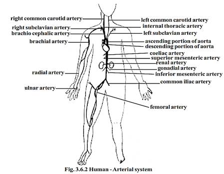
Components of Circulatory system Blood vessels
Blood vessels
The blood vessels carrying blood away from the heart are the arteries. The Veins carry blood towards the heart. The arteries and veins are named and classified according to their anatomical position. They can also be classified according to their size and wall structure. Functionally, arteries are subdivided into conducting, distributing and resistance vessels.
1-Conducting vessels: - These are large arteries from the heart and their main branches. The walls of these vessels are elastic in nature.
2-Distributing vessels: - These are smaller arteries reaching individual organs. They branch into the organs. They have muscular walls.
3-Resistance vessels: - These are mostly arterioles. While these vessels are smaller, their walls are highly muscular. Hence these vessels can reduce pressure of blood due to peripheral resistance.
4-Exchange vessels: - These are the capillaries. The walls of these vessels allow exchanges between blood and the tissue fluid surrounding the cells. The substances commonly exchanged are oxygen, carbon-di-oxide, nutrients, water, inorganic ions, vitamins, hormones, metabolic products and antibodies.
5-Capacitance or reservoir vessels:-These are the larger vessels and veins. These are of varying sizes. They collect and convey blood back to the heart. The higher capacitance of these vessels is due to their dispensability. Hence their blood content is more, even at low pressure. The number of such veins is also enormous. Thus the veins are called as the blood reservoirs""
Structure of blood vessels
The blood vessels show a vast range of structural modifications. However a few basic patterns can be studied.
A blood vessel consists of a wall and a lumen or cavity. The wall of the blood vessels is made up of 3 distinct layers or tunica. They are the tunica intima, tunica media and tunica externa or tunica adventitia.
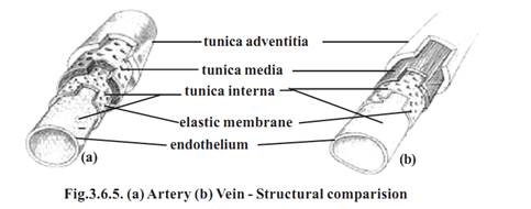
The tunica intima is formed of an endothelium, a delicate connective tissue and elastic fibres. The tunica media contains smooth muscle cells. It causes vasoconstriction and vasodilation. The tunica externa is composed of connective tissue. The composition and thickness of layers varies with the diameter of the blood vessels and the type.
Types of blood vessels
1-Large elastic arteries: - The walls of these arteries contain elastic fibres. The smooth wall measures about lmicron in thickness. It gets stretched under the effect of pulse and recoils elastically.
2-Muscular arteries: - There are larger and smaller muscular arteries. The larger muscular arteries are inelastic and they have thick walls. The wall has 30-40microns in diameter in the layers of smooth muscles. Since they regulate blood supply, they are called distributing arteries. The small muscular arteries are capable of vasodilation and vasoconstriction.
3-Arterioles: - They conduct blood from the arteries to the capillary bed. These are small vessels capable of vasodilation and vasoconstriction.
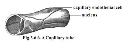
4-Capillaries: - These are fine vessels found between arterioles and venules. They measure 5-8micron in diameter.
5-Venules: - These are tubes of flat, oval or polygonal endothelial cells. Each venules is formed by the convergence of two or more capillaries. Its diameter ranges up to 30micron.
6-Veins: - Veins seen in anatomy are medium veins. They run in between venules and large veins. Large veins transport blood to the heart.
Veins with diameter above 2 mm have valves. They are of semilunar type. They allow movement of blood towards the heart. There are several valves in the medium veins
Branching of blood vessels: - When an artery divides into two equal branches, the original artery ceases to exist. Hence the branches are called terminal branches. The smaller branching vessels formed on the sides are called the collateral branches. When arteries are joined to each other it is named as anastomosis.
Blood supply to blood vessels: - As any other region, the cells and tissue on the wall of the blood vessel require nourishment. Some amount can diffuse from blood in the lumen. For vessels having diameter greater than 1 mm, diffusion of nutrients may not be possible. Such vessels have very minute vessels called vasa vasorum spread over them. They penetrate into the wall of the blood vessels.
Innervation of blood vessels: - The walls of the blood vessels are innervated by sympathetic nerve fibres. They regulate the contraction of the musculature. They effect vasoconstriction.
The Heart
The heart is a hollow, fibromuscular organ. It is somewhat conical or pyramidal in form. It is roughly the size of a closed fist. An average heart measures 12 cm from base to the apex. Transverse diameter at its broadest region is 8-9 cm. It is 6 cm thick antero-posteriorly. While in adult male the heart weighs 280-340 g, in female it weighs 230-280 g.
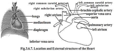
The thoracic organs such as heart, trachea and oesophagus form a midline partition called the mediastinum. The heart lies obliquely in the mediastinum.
The heart is surrounded by a double layered membrane called the pericardium. The outer layer is called the fibrous pericardium. The inner membrane is called the serous pericardium. In between heart and pericardium, there is a pericardial space. This space is filled with a fluid called the pericardial fluid.
The wall of the heart is made up of three tissue layers. They are the epicardium, myocardium and endocardium. The epicardium forms the smooth outer surface of the heart. The middle myocardium is composed of cardiac muscle. This layer plays an important role in the functioning of the heart. The endocardium forms the smooth inner surface. It is formed of squamous epithelium.
References
T. Sargunam Stephen, Biology (Zoology). First Edition – 2005, Government of Tamilnadu.


|
|
|
|
تفوقت في الاختبار على الجميع.. فاكهة "خارقة" في عالم التغذية
|
|
|
|
|
|
|
أمين عام أوبك: النفط الخام والغاز الطبيعي "هبة من الله"
|
|
|
|
|
|
|
قسم شؤون المعارف ينظم دورة عن آليات عمل الفهارس الفنية للموسوعات والكتب لملاكاته
|
|
|