آخر المواضيع المضافة

 النبات
الحيوان
الأحياء المجهرية
علم الأمراض
التقانة الإحيائية
التقنية الحيوية المكروبية
التقنية الحياتية النانوية
علم الأجنة
الأحياء الجزيئي
علم وظائف الأعضاء
الغدد
المضادات الحيوية
النبات
الحيوان
الأحياء المجهرية
علم الأمراض
التقانة الإحيائية
التقنية الحيوية المكروبية
التقنية الحياتية النانوية
علم الأجنة
الأحياء الجزيئي
علم وظائف الأعضاء
الغدد
المضادات الحيوية| Electron Microscopy |


|
|
Read More
Date: 26-10-2015
Date: 22-10-2015
Date: 26-10-2015
|
Electron Microscopy
The light microscope (LM) is limited in its resolution to about 0.25 mi- crometers. If two objects are closer together than that, they blur together and cannot be distinguished by the LM. The electron microscope (EM) overcomes this limitation and achieves resolutions down to 0.2 nanometers, allowing useful magnifications of biological material up to several hundred thousand times, and even more for nonbiological specimens. The EM achieves this by using a beam of electrons instead of visible light. Resolution is governed by the wavelength of illumination, and an electron beam has a much shorter wavelength (about 0.005 nanometers) than visible light (about 400 to 750 nanometers). Electron microscopes can therefore resolve objects as small as individual protein and deoxyribonucleic acid (DNA) molecules and pores in cell membranes.
The electron beam of an EM is generated by a heated tungsten wire (cathode) and accelerated down an evacuated column by a charge difference of typically 60,000 to 100,000 volts between the cathode and a grounded, mushroom-shaped anode. After passing through a hole in the center of the anode, it is focused on the specimen by electromagnets, which take the place of the glass lenses of a light microscope.
The Transmission Electron Microscope
In the transmission electron microscope (TEM), the electron beam passes through ultrathin tissue sections or small specimens, such as viruses. After passing through the specimen, the electrons strike a fluorescent screen and produce an image. The image can also be captured on photographic film or with a camera that digitizes it for storage on a computer.
Specimens for the TEM are typically fixed with aldehyde and stained with heavy metals, such as osmium, that will absorb or scatter electrons. The specimen is then dehydrated and embedded in a plastic resin. When it hardens, the resin is cut into sections 60 to 90 nanometers thick with a glass or diamond knife. Very tiny particles such as viruses and purified cell organelles can be viewed without sectioning by depositing them on a thin membrane. This membrane is treated with a heavy metal “negative stain” so that the specimen stands out as a light image against a dark background.
Areas of a specimen that bind the most osmium absorb the most energy from an electron beam, and are called electron-dense regions. Areas that bind less of the stain allow electrons to pass through more freely and are described as electron-lucent regions. Electrons that pass through the lightly stained, electron-lucent regions lose relatively little energy and produce relatively bright spots of light when they strike the screen. The more heavily stained, electron-dense regions cause some electrons to lose energy and others to be deflected from the beam, and thus produce dimmer spots on the screen. TEM images are essentially shadows caused by accumulations of the heavy metal on cellular structures or, in the case of negative staining, on the supporting membrane.
The Scanning Electron Microscope
The scanning electron microscope (SEM) is used to examine a specimen coated with vaporized metal ions (usually gold or palladium). An electron beam sweeps across the specimen surface and discharges secondary electrons from the metal coating. These electrons produce an image on a monitor similar to a television screen. The image on the monitor can be photographed or recorded with a digital camera. The SEM cannot see through a specimen as the TEM does, but can see only the surface where the metal coating is.
The SEM is capable of less resolution and useful magnification than the TEM. However, it produces dramatic three-dimensional images that can yield more information about surface topography than the flat images usually produced by TEM.
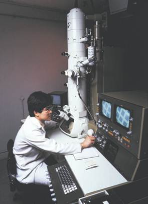
A scientist operating a scanning transmission electron microscope
Other Variations in Electron Microscopy
Both SEMs and TEMs can be equipped with a detector that monitors X rays given off by a specimen when it is bombarded by electrons. Other types of microscopes irradiate the specimen with ions or X rays and record ions, electrons, or X rays given off by the specimen. In both cases, the emitted particles and radiation yield information about the chemical composition of the specimen.
A scanning tunneling microscope measures the vertical movement of a tiny probe that is dragged over a specimen, producing a line representation of that movement. An atomic force microscope operates on a similar principle, but measures forces of attraction and repulsion between the specimen and the probe as the probe moves across the surface. In either case, multiple scan lines side by side produce images of the specimen surface, revealing details as small as the “atomic terrain” of individual molecules.
References
Berger, Dee. Journeys in Microspace: The Art of the Scanning Electron Microscope. New York: Columbia University Press, 1995.
Gilmore, C. P. The Scanning Electron Microscope: World of the Infinitely Small. Greenwich, NY: Graphic Society, 1972.
Microworld Internet Guide to Microscopy. <mwrn.com/guide/electron_microscopy/ microscope.htm>. Includes lecture notes and guides to EM techniques and instrumentation.
Slayter, Elizabeth M., and Henry S. Slayter. Light and Electron Microscopy. New York: Cambridge University Press, 1992.
WWW Virtual Library: Microscopy. <http://www.ou.edu/research/electron/mirror/>. Numerous links to other sites on all aspects of microscopy.


|
|
|
|
دخلت غرفة فنسيت ماذا تريد من داخلها.. خبير يفسر الحالة
|
|
|
|
|
|
|
ثورة طبية.. ابتكار أصغر جهاز لتنظيم ضربات القلب في العالم
|
|
|
|
|
|
|
قسم شؤون المعارف ووفد من جامعة البصرة يبحثان سبل تعزيز التعاون المشترك
|
|
|