آخر المواضيع المضافة

 النبات
الحيوان
الأحياء المجهرية
علم الأمراض
التقانة الإحيائية
التقنية الحيوية المكروبية
التقنية الحياتية النانوية
علم الأجنة
الأحياء الجزيئي
علم وظائف الأعضاء
الغدد
المضادات الحيوية
النبات
الحيوان
الأحياء المجهرية
علم الأمراض
التقانة الإحيائية
التقنية الحيوية المكروبية
التقنية الحياتية النانوية
علم الأجنة
الأحياء الجزيئي
علم وظائف الأعضاء
الغدد
المضادات الحيوية| Actin |


|
|
Read More
Date: 10-12-2015
Date: 31-12-2015
Date: 10-5-2016
|
Actin
Actin is one of the most ubiquitous and conserved eukaryotic proteins. While actin was originally isolated and studied as part of the contractile apparatus of muscle, we now know that actin is found in virtually all eukaryotic cells and can be the most abundant protein present. Actin forms part of the cytoskeleton in nonmuscle cells and is involved in the maintenance of cell shape as well as cell dynamics and motility. The active form of actin is a helical polymer called F-actin (F for filamentous), assembled from monomeric subunits of G-actin (G for globular). Actin exists in a number of very similar isoforms (88% amino acid identity between yeast cytoplasmic actin and human muscle actin), and these isoforms display tissue, rather than species, specificity (12, ).Thus, human cytoplasmic actin is much closer in sequence to yeast cytoplasmic actin than it is to human muscle actin. The actin monomer contains 374–376 residues, varying with the isoform, and is about 42k MW.
Actin interacts specifically with over 40 other proteins, and many of these interactions fall into separate classes. There are proteins that control the polymerization state of actin by nucleating, capping, or severing filaments [such as gelsolin, profilin, cofilin, and the b-thymosins (3-7)], proteins that crosslink actin filaments together into gels and bundles [such as a-actinin, villin, and fimbrin (8-10)], and proteins that regulate the interactions of myosin with actin [such as tropomyosin, troponin, calponin, and caldesmon (11), (see Thin Filament)]. In addition, many glycolytic enzymes bind actin, and it has been suggested that this provides spatial organization to metabolic processes via an association with the cytoskeleton (12). Other high-affinity interactions, such as between G-actin and the endonuclease DNase I (see DNase 1 Sensitivity) (13), do not lend themselves to simple explanations at this time.
Extensive biochemical, structural, and genetic studies have explored the properties of both G- and F-actin. The most detailed structural information exists for G-actin, since X-ray crystal structures have been obtained for complexes of G-actin with DNase I (13), gelsolin segment 1 (14), and profilin (15). The G-actin structure consists of two domains, each with two subdomains. These domains were originally called the “large” and “small” domains, but this terminology is not appropriate, as we now know that the “large” domain only contains 51% of the mass of the subunit. The N- and C-terminii are both located in the largest subdomain, subdomain-1. Between the two domains is a nucleotide-binding cleft, normally occupied by ATP in G-actin and ADP in F-actin, as well as the high-affinity metal-binding site. The subunit structure of G-actin is homologous to that of yeast hexokinase and the ATP-binding domain of HSC-70, a chaperonin, suggesting a common evolutionary origin for all of these proteins (16). It has been proposed that the bacterial protein FtsA has the same fold (17), which would indicate that FtsA is a prokaryotic ancestor of the eukaryotic actins. It has already been established that the bacterial FtsZ protein has a common structure with eukaryotic tubulins. Since neither hexokinase nor HSC-70 polymerize, it is evident that actin's ability to polymerize does not reside in any unique features of the secondary or tertiary structure, but rather evolved later. Thus, establishing that FtsA has a common subunit structure with actin will not, in and of itself, reveal any information about possible quaternary structures for FtsA.
Electron microscopic observations of F-actin (18-20) have been very useful in defining the orientation of the subunit in the filament, as well as revealing aspects of structural dynamics. X-ray fiber diffraction from oriented gels of F-actin has led to an atomic model for F-actin (21, 22), which provides a starting point for understanding the molecular details of the interactions of actin with many other proteins and ligands. However, many of the atomic details are still underdetermined by the method of fitting the monomeric structure into the filament, since the X-ray fiber diffraction pattern does not introduce all of the constraints needed. We thus have a general picture of the orientation of the subunit in the filament, but still lack information about many of the atomic interactions.
The subunit is oriented in the filament so that the two domains are nearly perpendicular to a plane containing the helix axis (Fig. 1). The resulting filament is about 100 Å in diameter, and subunits are spaced axially by a 27.3 Å rise along the filament axis. The change in the axial rise per subunit is less than .08 Å per subunit (<0.3%) when actin filaments in muscle are placed under full tension (23, (24, establishing that these filaments are relatively inextensible. Nevertheless, this amount of extensibility is enough to complicate the interpretation of muscle mechanics. Subunits are related to their axial neighbors by an ~167° rotation about the filament axis, giving rise to a 59 Å pitch left-handed helix (sometimes called the genetic helix). An interesting property of the actin filament is the ability of subunits to rotate within the helix (25), and it has recently been shown that an actin-binding protein, cofilin, can change the twist of the actin filament by 5° per subunit, without significantly changing the axial rise per subunit (7). The helical geometry of actin also gives rise to two right-handed long-pitch strands containing actin subunits related by a 54.6 Å axial separation. These strands typically have a mean pitch of ~700–760 Å (with the differences in the mean being due to the isoform, preparative conditions, associated proteins and ligands, etc.). The double-stranded character of these helices give rise to “crossovers” in projection at half of the pitch, or about 350–380 Å, but these crossover spacings can be variable due to the angular freedom within the actin filament (25, 26).
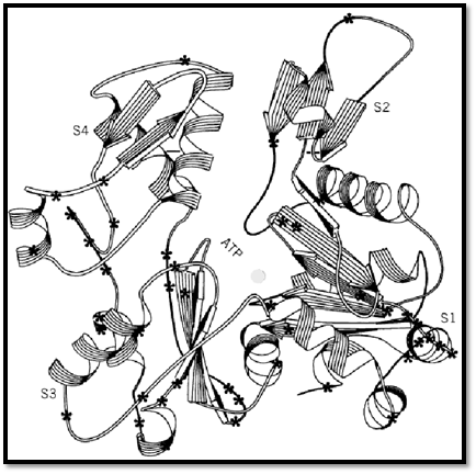
Figure 1. The G-actin subunit is shown by a ribbon representation (39) based upon the structure determined by X-ray crystallography (13). The degree of sequence conservation in actin is indicated by the fact that the only residues (45 out of 375) that differ between vertebrate striated skeletal muscle and yeast cytoplasmic actin are indicated by the asterisks. The four subdomains of actin are labeled S1–S4, and the most conserved part of the molecule is subdomain 2, with only 2 substitutions out of 38 residues between these two isoforms. The cleft between the two domains is occupied by ATP and a bound metal (indicated by the sphere). Adapted from Orlova et al. (40).
Every subunit in the actin filament is oriented with the same polarity, which gives rise to the overall polarity of the filament. This polarity has been described as the filament having a “barbed” end and a “pointed” end, based upon the morphological polarity that appears in electron micrographs when actin filaments are extensively decorated by the binding of proteolytic fragments of myosin, such as S1 or HMM (heavy meromyosin). This decoration generates a chevron appearance caused by all myosin molecules binding to the actin filament with an angle of about 45°. The morphological polarity has been related to a kinetic polarity apparent during in vitro polymerization, since the barbed end is the fast-growing end and the pointed end is the slow-growing end.
The helical symmetry of the actin filament is often described as 13/6 or 28/13, meaning that it has a helical repeat of 13 subunits in 6 turns of the 59 Å helix, or 28 subunits in 13 turns. The helical repeat is the distance that a subunit must be translated axially to bring it into register with another subunit. An ideal 13/6 helix would have crossovers every 355 Å, with each crossover containing a helical repeat, while an ideal 28/13 helix would have crossovers every 382 Å, with two crossovers per helical repeat. However, there is no reason to believe that the helical symmetry needs to be described as a ratio of small integers, as there is an infinitesimally small change in structure from a symmetry of 13/6 to one of 1301/600 (a rotation of ~0.1° per subunit), but the helical repeat changes from 355 Å to 35,517 Å! The actual symmetry of the actin filament is entirely defined by the interactions between one actin subunit and four nearest neighbors (–2 and 2 along the same long-pitch strand, and –1 and +1 on the opposite strand). In the absence of accessory proteins [such as tropomyosin, which binds to seven actin subunits along the same long-pitch strand (see Thin Filament)], all changes of state must be propagated by such local interactions. Nevertheless, many in vitro experimental observations have shown that pure actin (in the absence of accessory proteins) can have long-range cooperative interactions within a filament (20, 27-29).
The flexibility of the actin filament has been studied by many methods, including spectroscopy (30), light microscopy (31), and electron microscopy (32, 33). Most studies have suggested a persistence, or correlation, length of about 6–7 µm. This indicates that an actin filament much shorter than 6–7 µm can be treated as a rigid rod, while a filament much longer than 6–7 µm can be treated as a random coil. The persistence length does not suggest that a 6–7 µm filament is approximated well by a rigid rod, since an actin filament only 0.6 µm in length (one-tenth of the persistence length) will have a characteristic fluctuation of tangents at the two ends of about 25°.
Actin polymerization has been studied in vitro in great detail and is induced by raising the salt concentration. As for almost all other protein polymers in the cell, the assembly process involves the noncovalent binding of subunits to each other, such that every subunit (with the exception of subunits at the filament ends) is in the same environment. Probably more is known about the in vitro polymerization of actin than for any other protein polymer, but new findings suggest that the in vivo regulation of actin assembly involves the interactions with many other proteins and may be greatly different from what occurs in vitro (34). Polymerization can occur in vitro when the subunit concentration is above the critical concentration, which is the concentration of the monomer that would be at equilibrium with a population of polymers (see Treadmilling). Polymerization in vitro also requires that a monovalent salt, such as NaCl, be present at a concentration of ~100 mM, or a divalent salt such as MgCl2 be present at ~1 mM.
Actin has a single high-affinity metal binding site, which has a greater affinity for Ca2+ than for Mg2+. However, due to the much higher concentrations of Mg2+ than Ca2+ present in vitro, it is expected that this site will be occupied by Mg2+. This has been shown experimentally to be true in muscle (35), where the Mg2+ concentration is three orders of magnitude higher than the Ca2+ concentration. Actin also has 5–10 lower affinity metal binding sites (36). The spontaneous nucleation of actin filaments is often the rate-limiting step in actin polymerization in vitro, since the subsequent growth of existing filaments can occur rapidly. Within the cell, however, there is good reason to believe that actin polymerization occurs under the control of other proteins which serve to nucleate and cap growing filaments, and it is possible that spontaneous self-nucleation of actin filaments may never occur.
Actin polymerization is also loosely coupled to the hydrolysis of the bound nucleotide. While almost all attention has been focused on the primary nucleotide-binding site, actin also has a second nucleotide-binding site with mM affinity (37). Since this site is likely to be occupied in cells such as muscle where there exists a millimolar concentration of ATP, it remains to be seen whether this second site plays a physiological role. It is known that the nucleotide hydrolysis is not required for polymerization, since G-ADP actin can polymerize, as can G-actin subunits containing nonhydrolyzable analogs of ATP. The role of ATP hydrolysis is to actually destabilize the filament (38), allowing for depolymerization to occur more readily. ATP hydrolysis lags behind the initial polymerization event and occurs once a subunit is part of the polymer. The cell therefore uses the energy of ATP hydrolysis to allow for the dynamic assembly and disassembly of actin filaments, in contrast to less dynamic components of the cytoskeleton, such as intermediate filaments. The energy of ATP hydrolysis can also be used to drive treadmilling, where there is a unidirectional flux of subunits through a filament.
References
1. P. A. Rubenstein (1990) BioEssays 12, 309–315.
2. E. S. Hennessey, D. R. Drummond, and J. C. Sparrow (1993) Biochem. J. 291, 657–671.
3. E. Ballweber, E. Hannappel, T. Huff, and H. G. Mannherz (1997) Biochem. J. 327, 787–793.
4. M. F. Carlier and D. Pantaloni (1997) J. Mol. Biol. 269, 459–467.
5. D. A. Schafer and J. A. Cooper (1995) Ann. Rev. Cell Dev. Biol. 11, 497–518.
6. L. D. Burtnick, E. K. Koepf, J. Grimes, E. Y. Jones, D. I. Stuart, P. J. McLaughlin, and R. C. Robinson (1997) Cell 90, 661–670.
7. A. McGough, B. Pope, W. Chiu, and A. Weeds (1997) J. Cell Biol. 138, 771–781.
8. P. Matsudaira (1991) Trends Biochem. Sci. 16, 87–92.
9. D. Hanein et al. (1998) Nature Struct. Biol. 5, 787–792.
10. K. A. Taylor and D. W. Taylor (1994) Biophys. J. 67, 1976–1983.
11. L. S. Tobacman (1996) Ann. Rev. Physiol. 58, 447–481.
12. H. R. Knull and J. L. Walsh (1992) Curr. Top. Cell. Reg. 33, 15–30.
13. W. Kabsch, H. G. Mannherz, D. Suck, E. F. Pai, and H. C. Holmes (1990) Nature 347, 37–44.
14. P. J. McLaughlin, J. T. Gooch, H. G. Mannherz, and A. G. Weeds (1993) Nature 364, 685–692.
15. C. E. Schutt, J. C. Myslik, M. D. Rozycki, N. C. W. Goonesekere, and U. Lindberg (1993) Nature 365, 810–816.
16. K. M. Flaherty, D. B. McKay, W. Kabsch, and K. C. Holmes (1991) Proc. Natl. Acad. Sci. 88 5041 –5045 , .
17. P. Bork, C. Sander, and A. Valencia (1992) Proc. Natl. Acad. Sci. 89, 7290–7294.
18. R. A. Milligan, M. Whittaker, and D. Safer (1990) Nature 348, 217–221.
19. A. Orlova and E. H. Egelman (1995) J. Mol. Biol. 245, 582–597.
20. A. Orlova, E. Prochniewicz, and E. H. Egelman (1995) J. Mol. Biol. 245, 598–607.
21. K. C. Holmes, D. Popp, W. Gebhard, and W. Kabsch (1990) Nature 347, 44–49.
22. M. Lorenz, D. Popp, and K. C. Holmes (1993) J. Mol. Biol. 234, 826–836.
23. K. Wakabayashi, Y. Sugimoto, H. Tanaka, Y. Ueno, Y. Takezawa, and Y. Amemiya (1994) Biophys. J. 67, 2422–2435.
24. H. E. Huxley, A. Stewart, H. Sosa, and T. Irving (1994) Biophys. J. 67, 2411–2421.
25. E. H. Egelman, N. Francis, and D. J. DeRosier (1982) Nature 298, 131–135.
26. J. Hanson (1967) Nature 213, 353–356.
27. F. Oosawa (1983) In Muscle and Non-Muscle Motility (A. Stracher, ed.) Academic Press, New York, p. 151.
28. G. Drewes and H. Faulstich (1993) Eur. J. Biochem. 212, 247–253.
29. A. Muhlrad, P. Cheung, B. Phan, C. Miller, and E. Reisler (1994) J. Biol. Chem. 269, 1185211858 – .
30. T. Yanagida and F. Oosawa (1978) J. Mol. Biol. 126, 507–524.
31. H. Isambert, P. Venier, A. C. Maggs, A. Fattoum, R. Kassab, D. Pantaloni, and M. F. Carlier (1995) J. Biol. Chem. 270, 11437–11444.
32. T. Takebayashi, Y. Morita, and F. Oosawa (1977) Biochim. Biophys. Acta 492, 357–363.
33. A. Orlova and E. H. Egelman (1993) J. Mol. Biol. 232, 334–341.
34. M. F. Carlier (1998) Curr. Opin. Cell Biol. 10, 45–51.
35. T. Kitazawa, H. Shuman, and A. P. Somlyo (1982) J. Musc. Res. Cell Motil. 3, 437–454.
36. M. F. Carlier, D. Pantaloni, and E. D. Korn (1986) J. Biol. Chem. 261, 10778–10784.
37. P. Kiessling, B. Polzar, and H. G. Mannherz (1993) Biol. Chem. Hoppe-Seyler 374, 183–192.
38. C. Combeau and M. F. Carlier (1988) J. Biol. Chem. 263, 17429–17436.
39. P. J. Kraulis (1991) J. Appl. Crystallogr. 24, 946–950.
40. A. Orlova, X. Chen, P. A. Rubenstein, and E. H. Egelman (1997) J. Mol. Biol. 271, 235–243.


|
|
|
|
دخلت غرفة فنسيت ماذا تريد من داخلها.. خبير يفسر الحالة
|
|
|
|
|
|
|
ثورة طبية.. ابتكار أصغر جهاز لتنظيم ضربات القلب في العالم
|
|
|
|
|
|
|
أصواتٌ قرآنية واعدة .. أكثر من 80 برعماً يشارك في المحفل القرآني الرمضاني بالصحن الحيدري الشريف
|
|
|