آخر المواضيع المضافة

 النبات
الحيوان
الأحياء المجهرية
علم الأمراض
التقانة الإحيائية
التقنية الحيوية المكروبية
التقنية الحياتية النانوية
علم الأجنة
الأحياء الجزيئي
علم وظائف الأعضاء
الغدد
المضادات الحيوية
النبات
الحيوان
الأحياء المجهرية
علم الأمراض
التقانة الإحيائية
التقنية الحيوية المكروبية
التقنية الحياتية النانوية
علم الأجنة
الأحياء الجزيئي
علم وظائف الأعضاء
الغدد
المضادات الحيوية| Laboratory Diagnosis of virus |


|
|
Read More
Date: 19-11-2015
Date: 2025-02-22
Date: 19-11-2015
|
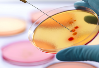
Laboratory Diagnosis of virus
The following methods can be used to obtain a virological laboratory diagnosis:
-Virus isolation by growing the pathogen in a compatible host; usually done in cell cultures, rarely in experimental animals or hen embryos.
-Direct virus detection. The methods of serology, molecular biology, and electron microscopy are used to identify viruses or virus components directly, i.e., without preculturing, in diagnostic specimens.
-Serodiagnostics involving assay of antiviral antibodies of the IgG or IgM classes in patient serum.
Indication and methods. Laboratory diagnostic procedures for virus infections are costly, time-consuming, and require considerable staff time. It is therefore important to consider carefully whether such tests are indicated in a confirmed case. The physician in charge of treatment must make this decision based on detailed considerations. In general, it can be said that laboratory diagnostics are justified if further treatment of the patient would be influenced by an etiological diagnosis or if accurate diagnostic information is required in the context of an epidemic or scientific research and studies.
There are essentially three different methods used in virological diagnostics (Table 1):
1 .Virus isolation by growing the pathogen in a compatible host; usually done in cell cultures.
2 .Direct virus detection in patient material; identification of viral particles using electron microscopy, viral antigens with the methods of serology, and viral genome (components) using the methods of molecular biology.
3 .Antibody assay in patient serum.
General guidelines for viral diagnostics are listed below.
Virus Isolation by Culturing
In this approach, the virus is identified based on its infectivity and pathogenicity by inoculating a host susceptible for the suspected virus—in most cases cell cultures—with the specimen material. Certain changes observed in the culture (cytopathic effect [CPE]) indicate the presence of avirus.
Sampling and transport of diagnostic specimens. Selection of suitable material depends on the disease and suspected viral species (see Chapter 8). Sampling should generally be done as early as possible in the infection cycle since, as was mentioned on viral replication precedes the clinical symptoms. Sufficiently large specimens must be taken under conditions that are as sterile as possible, since virus counts in the diagnostic material are almost always quite low. Transport must be arranged quickly and under cold box conditions. The half-life of viruses outside the body is often very short and must be extended by putting the material on ice. A number of virus transport mediums are commercially available. A particular transport medium should be selected after consulting the laboratory to make sure the medium is compatible with the laboratory methods employed. Such mediums are particularly important if the diagnostic material might otherwise dry out.
Information provided to the laboratory. The laboratory must be provided with sufficient information concerning the course and stage of the disease, etc. This is very important if the diagnostic procedure is to be efficient and the results accurate. Clinical data and tentative diagnoses must be provided so the relevant viruses can be looked for in the laboratory. Searching for every single virus potentially present in the diagnostic material is simply not feasible for reasons of cost and efficiency.
Laboratory processing of the material. Before the host is inoculated with the specimen material for culturing, contaminant bacteria must be eliminated with antibiotics, centrifugation, and sometimes filtering. All of these manipulations of course entail the risk of virus loss and reduction of test sensitivity, so the importance of sterile sampling cannot be overemphasized. In a few cases, virus enrichment is indicated, e.g., by means of ultracentrifugation.
Selection of a host system. The host system to be used is chosen based on the suspected (and relevant) virus infectors. Observation and incubation times, and thus how long a laboratory diagnosis will take, also depend on the viral species under investigation.
Identification of the viruses is based first on the observed cell changes, then determined serologically using known antibodies and appropriate methods such as immunoelectron microscopy, EIA, or the neutralization test. Methods that detect the viral genome by means of in-situ or filter hybridization are now seeing increasing use.
Significance of results. The importance of virus isolation depends on the virus type. In most cases, isolation will be indicative of the etiology of the patient's disease. In some cases, (in particular the herpesvirus and adenovirus group, see Chapter 8), latent viruses may have been activated by a completely different disease. In such cases, they may of course be isolated, but have no causal connection with the observed illness.
Isolation is the most sensitive method of viral diagnostic detection, but it cannot detect all viruses in all situations. This means that a negative result does not entirely exclude a viral infection. Another aspect is that the methods of virus isolation, with few exceptions, detect only mature, infectious virions and not the latent viruses integrated in the cells. This renders diagnostic isolation useless during latency (e.g., herpes simplex between recidivations).
Amplification culture. In this method, the virus is grown for a brief period in a cell culture. Before the CPE is observed, the culture is tested using the antigen and genomic methods described. This is also known as a “shell vial assay" because the cells are grown on coverslips in shell vials (test tubes with screw caps). Using this arrangement, method sensitivity can be increased by centrifuging the diagnostic material onto the cell monolayer. The greatest amount of time is saved by detecting the virus-specific proteins produced early in the infection cycle, which is why the search concentrates on such so-called “early antigens” (see p. 388). Using this method, the time required to confirm a cytomegaly virus, for instance, can be shortened from four to six weeks to only two to five days with practically no loss of sensitivity compared to classic isolation methods.
Direct Virus Detection
In this diagnostic approach, the viruses are not identified as infectious units per se, but rather as viral particles or parts of them. The idea is to find the viruses directly in the patient material without prior culturing or replication. Viruses in serous fluids such as the contents of herpes simplex or varicellazoster blisters can be viewed under the electron microscope (EM). It must be remembered, however, that the EM is less sensitive than virus isolation in cultures by a factor of 105. Viral antigens can be detected in secretions using enzyme immunoassay (EIA), passive agglutination, or in smears with immunofluorescence performed with known antibodies, for instance monoclonal antibodies. Analogously, the viral genome can be identified by means of filter hybridization, or in smears or tissue sections with in-situ hybridization using DNA or RNA complementary to the viral genome as a probe.
Sampling and transport of diagnostic specimens. Transport of patient material for these methods is less critical than for virus isolation. Cold box transport is usually not required since the virus need not remain infectious.
-Electron microscopy. For negative contrast EM, the specimen is transported to the laboratory without any additives (dilution!).
-Antigen assay. For an immunofluorescence antigen assay, slide preparations must be made and fixed immediately after sampling. Special extraction mediums are used in EIA. Since commercial kits are used in most cases, procedure and reagents should be correlated with the laboratory.
-Genome hybridization. Here as well, the specimen material must meet specific conditions depending on whether the viruses are to be identified by the in-situ method or after extraction. This must be arranged beforehand with the laboratory.
Significance of results. A positive result with a direct virus detection method has the same level of significance as virus isolation. A negative test result means very little, particularly with EM, due to the low level of sensitivity of this method. The antigen assay and genome hybridization procedures are more sensitive than EM, but they are selective and detect only the viruses against which the antibodies or the nucleic acid probe used, are directed. It is therefore of decisive importance to provide the laboratory with detailed information.
Virus Detection Following Biochemical Amplification
Polymerase chain reaction (PCR, Fig. 1). This method provides a highly sensitive test for viral genomes. First, nucleic acid is extracted from the patient material to be analyzed. Any RNA virus genome present in the material is transcribed into DNA by reverse transcriptase). This DNA, as well as the DNA of the DNA viruses, is then replicated in vitro with a DNA polymerase as follows: after the DNA double strand has been separated by applying heat, two synthetic oligonucleotides are added that are complementary to the two ends of the viral genome segment being looked for and can hybridize to it accordingly. The adjacent DNA (toward each 5' end) is then copied with an added polymerase, whereby

Fig. 1 a Two oligonucleotide primers are hybridized to the DNA double strands, which have been separated by heating. A heat-stable polymerase (e.g., Taq polymerase from Thermus aquaticus) is then added and extends these primers along the length of, and complementary to, the matrix strand. The resulting double strands are then once again separated by heat and the reaction is repeated. b The DNA strands produced in the first cycle (1st generation) have a defined 5' end (corresponding to the primer) and an undefined 3' end. All of the subsequent daughter strands (2nd to nth generation) have a uniform, defined length.
the oligonucleotides act as primers. The new and old strands are once again separated by heat and the reaction is started over again. Running several such cycles amplifies the original viral DNA by a factor of many thousands. Beginning with the second generation, the newly synthesized DNA strands show a uniform, defined length and are therefore detectable by means of gel electrophoresis. The specificity of the reaction is verified by checking the sequences of these DNA strands by means of hybridization or sequencing. The amplification and detection systems in use today for many viruses are increasingly commercially available, and in some cases are also designed to provide quantitative data on the “viral load.”
Serodiagnosis
If a viral infection induces humoral immunity, the resulting antibodies can be used in a serodiagnosis. When interpreting the serological data, one is confronted by the problem of deciding whether the observed reactions indicate a fresh, current infection or earlier contact with the virus in question. Two criteria can help with this decision:
Detection of IgM (without IgG) proves the presence of a fresh primary infection. IgM is now usually detected by specific serum against human IgM in the so-called capture test, an EIA.
To test for IgM alone, a blood specimen must be obtained very early in the infection cycle. Concurrent detection of IgG and IgM in blood sampled somewhat later in the course of the disease would also indicate a fresh infection. It could, however, also indicate a reactivated latent infection or an anamnestic reaction (i.e., a nonspecific increase in antibodies in reaction to a nonrelated infection), since IgM can also be produced in both of these cases.
A fourfold increase in the IgG titer within 10-14 days early on in the course of the infection or a drop of the same dimensions later in the course would also be confirmation.
References
Fritz H. Kayser, M.D. Emeritus Professor of Medical Microbiology Institute of Medical Microbiology, University of Zurich, Zurich, SwitzerlandThieme 2005, Stuttgart ! New York.


|
|
|
|
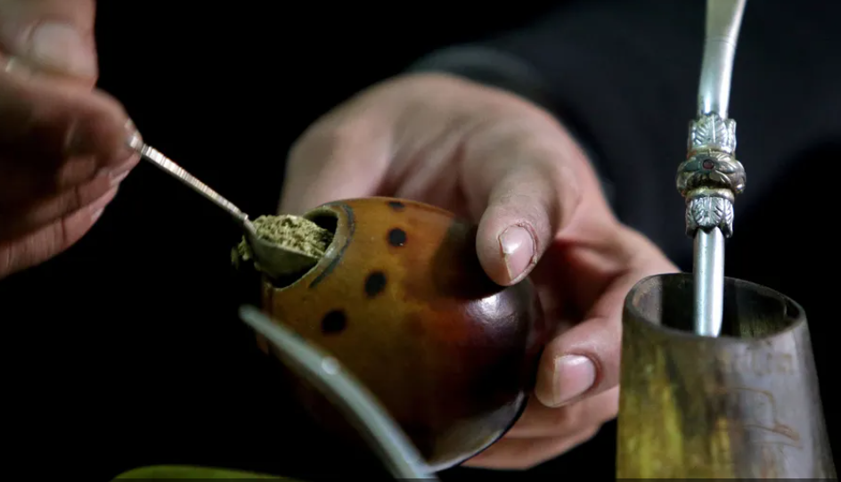
"إنقاص الوزن".. مشروب تقليدي قد يتفوق على حقن "أوزيمبيك"
|
|
|
|
|
|
|
الصين تحقق اختراقا بطائرة مسيرة مزودة بالذكاء الاصطناعي
|
|
|
|
|
|
|
قسم شؤون المعارف ووفد من جامعة البصرة يبحثان سبل تعزيز التعاون المشترك
|
|
|