آخر المواضيع المضافة

 النبات
الحيوان
الأحياء المجهرية
علم الأمراض
التقانة الإحيائية
التقنية الحيوية المكروبية
التقنية الحياتية النانوية
علم الأجنة
الأحياء الجزيئي
علم وظائف الأعضاء
الغدد
المضادات الحيوية
النبات
الحيوان
الأحياء المجهرية
علم الأمراض
التقانة الإحيائية
التقنية الحيوية المكروبية
التقنية الحياتية النانوية
علم الأجنة
الأحياء الجزيئي
علم وظائف الأعضاء
الغدد
المضادات الحيوية| Respiratory system |


|
|
Read More
Date: 16-7-2021
Date: 21-7-2021
Date: 21-7-2021
|
Respiratory system
The process of respiration involves movement of air in and out of the lungs, gas exchange between air in lungs and the blood, transport of O2 and CO2. These processes are facilitated by working together of well-developed respiratory organs and the circulatory system.
The respiratory organs include nasal cavity, pharynx, larynx, trachea, bronchi and lungs. These organs are organized into upper and lower respiratory tracts.

1-Nasal cavity: The nasal cavity follows the external nose. The nose is a visible prominent structure. Internally it is supported by cartilage plates. The bridge of the nose is formed of the nasal bones and extension of the skull bones (frontal and maxillary). The respiratory passage is divided into two chambers by a median partition. The nasal passage opens to the outside through external nostrils. It opens inside by internal nostrils at the pharynx.
2-Pharynx: The buccal cavity and the nasal passage open into the pharynx. It is a common pathway that opens into the oesophagus of the alimentary canal and larynx of the respiratory system.
The pharynx is divided into three regions, namely the nasopharynx, the oropharynx and the laryngopharynx.
The nasopharynx extends from the internal nostril to the region of the uvula. The uvula is a soft outgrowth hanging in between the posterior part of the oral cavity and the pharynx. It prevents the entry of food into the nasal cavity. The wall of the nasopharynx is lined by ciliated columnar epithelium. The middle ear opens into the nasopharynx through two auditory tubes. This arrangement is meant for equalizing the air pressure between the atmosphere and the middle ear. The inner surface of the nasopharynx also contains the pharyngeal tonsil or adenoid meant for defense against infections. An enlargement of the tonsil can interfere with breathing.
The oropharynx remains between the uvula and the epiglottis. The oral cavity opens into the oropharynx. Near the opening of the oral cavity 2 sets of palatine tonsils and lingual tonsils are present.
The laryngopharynx extends in between the epiglottis and the oesophagus.
3-Larynx: The larynx is seen just behind the pharynx and the buccal cavity. This region is surrounded by cartilages (3 unpaired and 6 paired). These are interconnected by muscles and ligaments.
The unpaired cartilages are the thyroid, cricoid and epiglottis. The thyroid cartilage is the largest. It is also known as the Adam’s apple.

The cricoid cartilage forms the base of the larynx. The other cartilages are placed above the cricoid. The epiglottis is attached to the thyroid. It projects as a free flap over the opening of the larynx. It prevents food particles from saentering into the tracheal tube.
The ligaments inside the larynx form the vocal folds or vocal cords. The vocal cords and the openings between them are called the glottis. The vocal cords are involved with sound production. The air moving past the vocal cords make them to vibrate. Louder sounds are made by increasing the amplitude of vibrations. Frequency of the vibrations can be altered by changing the length of the vibrating segments of the vocal cords. The length is altered by muscles attached to the cartilage. Males usually have longer vocal cords than females. The sound made by the vocal cords can be altered by the tongue, lips and teeth to form words.
4-Trachea (or wind pipe): It is a membranous tube. The wall is made up of connective tissue and smooth muscles. The wall is provided support by 15-20 ‘C’ shaped cartilage rings. They protect the trachea and keep it open all the time.
The inner wall of the trachea is lined by mucous membrane. It consists of ciliated columnar epithelium. The cilia of this epithelium help to propel mucus and foreign particles towards the larynx.
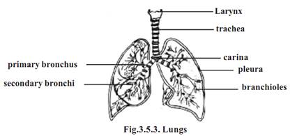
The length of the trachea is 10-12 cm. Its inner diameter is 12 mm. The trachea extends from the larynx to the level of the 5th thoracic vertebra. The basal part of the trachea divides to form 2 smaller tubes called the primary bronchi (sing: bronchus). The cartilage ring found at the basal region is called the carina. Foreign objects reaching carina stimulate a powerful cough reflex.
5-Lungs: the pair of lungs is the actual organs of respiration. Each lung is conical in shape. The base of the lung rests on the diaphragm. The right lung is larger than the left and it weighs around 620g. The left lung weighs 560g. The right lung has three lobes and the left lung has two.
The lungs are placed within the thoracic cavity. Each lung is surrounded by separate pleural membrane. The region inside the pleural membrane is named as the pleural cavity. This cavity is filled with pleural fluid.
The region in between the two lungs is named as the mediastinum. It is a midline partition, being occupied by the heart, trachea and oesophagus.
Structures such as the primary bronchi, blood vessels, nerves and lymphatic vessels enter or exit the lungs at a specific region on the inner margin of the lungs. This region is known as the hilum. All structures passing through the hilum are referred to as the root of the lung.
The primary bronchi on entering into each lung divide further into secondary bronchi. There are two secondary bronchi in the left lung and three in the right lung. The secondary bronchi in turn give rise to tertiary bronchi. They divide still further and finally give rise to bronchioles. The diameter of the bronchioles is less than 1 mm. These bronchioles divide several times to become still smaller terminal bronchioles.
Like the trachea, the primary bronchi are supported by ‘C’. Shaped cartilages and smooth muscles. As the bronchi become smaller the cartilages are replaced by smooth muscles.
The terminal bronchioles end in small air filled chambers called alveoli. The alveoli are thin walled pouches. They collectively provide the respiratory surface for gaseous exchange. The wall of the alveolus is very thin providing a minimal barrier to gaseous exchange between air and blood. The thickness of the wall of the alveolus is as little as 0.05m. Studies have shown that in human lungs there are about 300 million alveoli. They provide a mean total alveolar surface area value of 143 m2.
6-Thoracic wall and muscles of respirations
Eventhough the lungs are the principal organs of respiration, the process of ventilation happens by an indirect method. Air pressure gradients between thoracic chamber and lung cavity due to thoracic enlargement and reduction cause ventilation of lungs. Thoracic modifications during respiration happen due to several muscles. These are called the muscles of inspiration and expiration. These muscles are the diaphragm, external and internal intercostal muscles between the ribs, pectorals and scalene (Ref. Muscular system).
References
T. Sargunam Stephen, Biology (Zoology). First Edition – 2005, Government of Tamilnadu.


|
|
|
|
إدارة الغذاء والدواء الأميركية تقرّ عقارا جديدا للألزهايمر
|
|
|
|
|
|
|
شراء وقود الطائرات المستدام.. "الدفع" من جيب المسافر
|
|
|
|
|
|
|
العتبة العبّاسيّة: البحوث الّتي نوقشت في أسبوع الإمامة استطاعت أن تثري المشهد الثّقافي
|
|
|