آخر المواضيع المضافة

 النبات
الحيوان
الأحياء المجهرية
علم الأمراض
التقانة الإحيائية
التقنية الحيوية المكروبية
التقنية الحياتية النانوية
علم الأجنة
الأحياء الجزيئي
علم وظائف الأعضاء
الغدد
المضادات الحيوية
النبات
الحيوان
الأحياء المجهرية
علم الأمراض
التقانة الإحيائية
التقنية الحيوية المكروبية
التقنية الحياتية النانوية
علم الأجنة
الأحياء الجزيئي
علم وظائف الأعضاء
الغدد
المضادات الحيوية| Spinal Cord |


|
|
Read More
Date: 9-7-2021
Date: 21-7-2021
Date: 16-7-2021
|
Spinal Cord
Housed and protected within the vertebral column, the spinal cord spans from the foramen magnum to approximately the L2 vertebral level in the adult (Fig. 1). As part of the central nervous system (CNS), the spinal cord serves as a reflex center and conducts motor and sensory signals between the brain and the body. The vertebral column also contains vasculature, adipose, and meningeal layers, which supply and protect the spinal cord, respectively. The spinal cord is cylindrical in shape with enlargements in the cervical and lumbar regions to accommodate for upper and lower limb innervation, respectively. Distally, the spinal cord tapers and ends at L2 as the conus medullaris. Multiple spinal nerve rootlets emerge from the distal spinal cord and conus medullaris to exit the vertebral column. They are collectively referred to as the cauda equina .
A. Internal organization
As shown in Figure 2, in cross section, the spinal cord has an "H"-shaped gray matter core made up of neuronal cell bodies and peripheral white matter made up of nerve fibers (axons).
1. Gray matter: Gray matter is organized into anterior and posterior horns, which contain motor and sensory cell bodies, respectively. The thoracolumbar region {T1-L/L3) also features a lateral horn that contains preganglionic sympathetic cell bodies organized into an intermediolateral cell column.
2. White matter: Peripheral white matter is organized into specific ascending (sensory) and descending (motor) tracts.
B. Spinal nerves
Spinal nerves carry autonomic and somatic motor (general visceral efferent [GVE] and general somatic efferent [GSE]) and sensory (general visceral afferent [GVA] and general somatic afferent [GSA]) nerve fibers. Thirty-one paired spinal nerves exit the vertebral column through the intervertebral foramina or sacral foramina at each vertebral level (8 cervical, 12 thoracic, 5 lumbar, 5 sacral, and 1 coccygeal).

Figure 1:Spinal cord. Posterior schematic image of the spinal cord within the vertebral column (laminae removed) and exiting spinal nerves.

Figure 2: Cross section of the spinal cord with spinal nerve components.
1. Anterior and posterior rootlets: These arise from the spinal cord laterally, representing motor and sensory fibers, respectively (see Fig. 2). Rootlets converge to form nerve roots, which contain autonomic and somatic motor (anterior root) or sensory (posterior root) components.
2. Pseudounipolar sensory neurons: Cell bodies for these are housed in the spinal ganglion (posterior root ganglion) that is contained in the posterior root (Fig. 3).
3. Multipolar motor neurons: Cell bodies for these are found in the anterior horn of the spinal cord, and their axons form the anterior root.
4. Rami: Once the spinal nerve has exited the vertebral column, it splits into an anterior and a posterior ramus. Anterior rami provide motor and sensory innervation to the skin, muscles, vasculature, and joints of the trunk and extremities, while the posterior rami innervate intrinsic back muscles, associated vasculature, and overlying skin.

Figure 3 :Cadaveric image showing caudal spinal cord structures {laminae removed).
C. Meninges
Spinal meninges include the dura mater, arachnoid mater, and pia mater, which collectively surround, support, and protect the spinal cord and spinal nerve roots (Fig. 4).
1. Dura mater: The dura mater (Latin for tough mother) is the outermost layer and is a thick, tough covering. An epidural (extradural) space separates the dura from the boundaries of the vertebral foramen that is filled with adipose and a spinal venous plexus. Spinal dura mater is continuous with the cranial dura mater and extends inferiorly to the level of S2, forming a tube-like dural sac (Fig. 4).
2. Arachnoid mater: Deep to the dura mater is the arachnoid mater, a delicate, web-like layer that creates a subarachnoid space filled with cerebrospinal fluid (CSF). CSF bathes and protects the spinal cord and nerve roots within the vertebral canal. The subarachnoid space also contains arterial and venous branches that nourish the spinal cord. Under normal conditions, the arachnoid and dura are closely associated, obliterating the subdural space between these two layers. However, bleeding can occur into this space, forming a subdural hematoma. A widened subarachnoid space exists below the level of the conus medullaris between vertebral levels L2-S2. This space, referred to as the lumbar cistern, lies within the dural sac and contains CSF and the cauda equina.
3. Pia mater: The innermost meningeal layer, the pia mater, is closely adhered to the spinal cord. Two specializations of pia mater function to anchor the spinal cord within the vertebral canal: Denticulate ligaments are extensions of pia that project laterally at each vertebral foramen level and anchor into the inner surface of the dura mater, separating anterior and posterior rootlets (Fig. 5). The filum terminale is an extension of pia from the tip of the conus medullaris that extends inferiorly to attach on the coccyx, acting as a caudal anchor for the spinal cord.
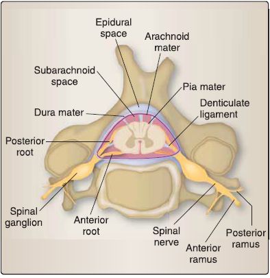
Figure4 : Cross-sectional view of spinal meninges and spaces.

Figure 5 : Lumbosacral myelography.


|
|
|
|
للعاملين في الليل.. حيلة صحية تجنبكم خطر هذا النوع من العمل
|
|
|
|
|
|
|
"ناسا" تحتفي برائد الفضاء السوفياتي يوري غاغارين
|
|
|
|
|
|
|
نحو شراكة وطنية متكاملة.. الأمين العام للعتبة الحسينية يبحث مع وكيل وزارة الخارجية آفاق التعاون المؤسسي
|
|
|