آخر المواضيع المضافة

 النبات
الحيوان
الأحياء المجهرية
علم الأمراض
التقانة الإحيائية
التقنية الحيوية المكروبية
التقنية الحياتية النانوية
علم الأجنة
الأحياء الجزيئي
علم وظائف الأعضاء
الغدد
المضادات الحيوية
النبات
الحيوان
الأحياء المجهرية
علم الأمراض
التقانة الإحيائية
التقنية الحيوية المكروبية
التقنية الحياتية النانوية
علم الأجنة
الأحياء الجزيئي
علم وظائف الأعضاء
الغدد
المضادات الحيوية| Great Saphenous Vein and Vena Cava Inferior |


|
|
Read More
Date: 25-7-2016
Date: 14-8-2016
Date: 5-1-2017
|
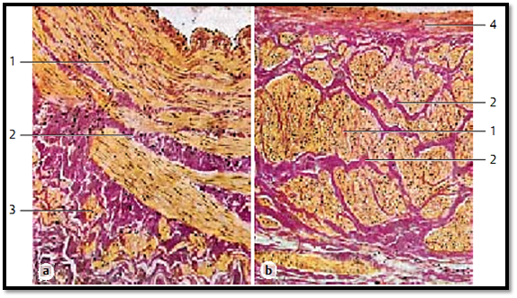
Great Saphenous Vein and Vena Cava Inferior
The two cross-sections show the vast differences in the ways the structural units are arrange d in the venous walls of the great saphenous vein (a) and the inferior vena cava (b). At the same time, the sections give an impression about the presence of muscle tissue (yellow ) and connective tissue (re d) in the large venous walls of the lower body. While the muscle cell bundles of the tunica me dia of the great saphenous vein 1 show a more or less circular arrangement, the muscle cell bundles of the tunica me dia of the lower central vein (vena cava) (b) show a longitudinal orientation. Strong septa of collagenous connective tissue 2 separate the muscle cell bundles. In both cases, the tunica interna is locate d at the top. Note the muscle bundles (yellow) in the tunica externa of the great saphenous vein 3 . The inferior vena cava shows a wider connective tissue layer 4 underneath the tunica intima.
1 Tunica media
2 Connective tissue septa
3 Tunica externa
4 Lamina propria intimae
Stain: van Gieson iron hematoxylin-picrofuchsin; magnification: × 15
References
Kuehnel, W.(2003). Color Atlas of Cytology, Histology, and Microscopic Anatomy. 4th edition . Institute of Anatomy Universitätzu Luebeck Luebeck, Germany . Thieme Stuttgart · New York .


|
|
|
|
"عادة ليلية" قد تكون المفتاح للوقاية من الخرف
|
|
|
|
|
|
|
ممتص الصدمات: طريقة عمله وأهميته وأبرز علامات تلفه
|
|
|
|
|
|
|
المجمع العلمي للقرآن الكريم يقيم جلسة حوارية لطلبة جامعة الكوفة
|
|
|