آخر المواضيع المضافة

 النبات
الحيوان
الأحياء المجهرية
علم الأمراض
التقانة الإحيائية
التقنية الحيوية المكروبية
التقنية الحياتية النانوية
علم الأجنة
الأحياء الجزيئي
علم وظائف الأعضاء
الغدد
المضادات الحيوية
النبات
الحيوان
الأحياء المجهرية
علم الأمراض
التقانة الإحيائية
التقنية الحيوية المكروبية
التقنية الحياتية النانوية
علم الأجنة
الأحياء الجزيئي
علم وظائف الأعضاء
الغدد
المضادات الحيوية| Membranous Osteogenesis-Parietal Bone |


|
|
Read More
Date: 23-1-2017
Date: 28-7-2016
Date: 25-7-2016
|
Membranous Osteogenesis-Parietal Bone
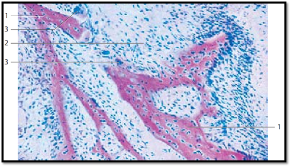
Membranous osteogenesis (intramembranous ossification) creates connective tissue bones ( direct osteogenesis). During direct osteogenesis, mesenchyme cells in the embryonic connective tissue differentiate into osteoblasts (initiation of ossification). Osteoblasts are rich in ergastoplasm and secrete an amorphous ground substance, which contains glycoproteins and proteoglycans as well as the precursors of collagen fibrils. Membranous osteogenesis is therefore always accompanied by the appearance of a matte d network of very delicate fibrils. Osteoblasts form a soft bone precursor material, which is termed osteoid bone. This figure shows red-stained osteoid lamella 1 . Large osteoblasts line up at their surface. The osteoid lamellae grow by apposition. The cells inside the osteoid lamellae have entrapped themselves during osteogenesis and have differentiated into osteocytes in the process. The connective tissue between the lamellae 2 is rich in cells. In the left half of the figure, there are osteoid lamellae in the immediate vicinity of multinucleated giant cells. These are osteoclasts 3 , which degrade bone tissue.
1 Osteoid lamellae with osteocytes 3 Osteocytes
2 Embryonic connective tissue
Stain: hematoxylin-eosin; magnification: × 90
Membranous Osteogenesis—Parietal Bone
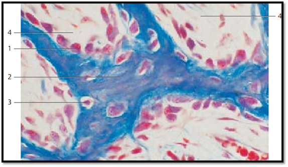
Genesis of connective tissue-derived bone in the area of the parietal bone of a human fetus. The osteoid lamella is surrounded by many osteoblasts 1 . They form prebone tissue, the osteoid 2 (here stained blue) and contain osteocytes 3 . A fibrous structure is still discernible (cartilage bone, fibrous bone).
The richly vascularized mesenchymal tissue between the primary bony lamellae (spongy bone trabeculae) is called primary bone marrow 4 . Later, secondary bone marrow, which forms the blood cells, will arise from it.
1 Osteoblasts 3 Osteocytes
2 Osteoid with fiber s 4 Primary bone marrow
Stain: azan; magnification: × 400
Membranous Osteogenesis—Parietal Bone
Multinucleated giant cells, osteoclasts, soon appear on the scenes of osteogenesis. They are part of the hematopoietic macrophage-monocyte system and are able to systematically degrade the basic bone substance (bone resorption) and phagocytose the products of this degradation. Osteoblasts have irregular shapes. They are branched and may contain as many as 50 or more nuclei. Osteoblasts are located close to the newly formed osteoid lamellae. When they degrade osteoid material, they create small bays, termed How-ship lacunae or arrosion bays (lacunar resorption). Only a few nuclei are visible in the figure. However, it provides an image of the lacy projections of the osteoclasts. In the lower part of the figure is an osteoid lamella. The area right and left of the osteoclast represents primary bone marrow.
Stain: hematoxylin-eosin; magnification: × 400

References
Kuehnel, W.(2003). Color Atlas of Cytology, Histology, and Microscopic Anatomy. 4th edition . Institute of Anatomy Universitätzu Luebeck Luebeck, Germany . Thieme Stuttgart · New York .


|
|
|
|
دخلت غرفة فنسيت ماذا تريد من داخلها.. خبير يفسر الحالة
|
|
|
|
|
|
|
ثورة طبية.. ابتكار أصغر جهاز لتنظيم ضربات القلب في العالم
|
|
|
|
|
|
|
العتبة العباسية المقدسة تستعد لإطلاق الحفل المركزي لتخرج طلبة الجامعات العراقية
|
|
|