آخر المواضيع المضافة

 النبات
الحيوان
الأحياء المجهرية
علم الأمراض
التقانة الإحيائية
التقنية الحيوية المكروبية
التقنية الحياتية النانوية
علم الأجنة
الأحياء الجزيئي
علم وظائف الأعضاء
الغدد
المضادات الحيوية
النبات
الحيوان
الأحياء المجهرية
علم الأمراض
التقانة الإحيائية
التقنية الحيوية المكروبية
التقنية الحياتية النانوية
علم الأجنة
الأحياء الجزيئي
علم وظائف الأعضاء
الغدد
المضادات الحيوية| Leptospira |


|
|
Read More
Date: 8-3-2016
Date: 15-3-2016
Date: 14-3-2016
|
Leptospira (Leptospirosis, Weil Disease)
The pathogenic species Leptospira interrogans is subclassified in over 100 serovars reflecting different surface antigens. The serovars are divided into 19 sero-groups (icterohemorrhagiae, canicola, pomona, etc.). These organisms are in the form of spiral rods and can be grown in in-vitro cultures. Leptospirosis is a zoonosis that occurs worldwide. The sole sources of infection are diseased rodents and domestic animals (pigs), which excrete the pathogen in their urine. Upon contact, leptospirae penetrate skin or mucosa, are disseminated hematogenously and cause a generalized vasculitis in various organs. The incubation period is seven to 12 days. The disease at first presents as a sepsis, followed after three to seven days by the so-called immune stage. In the milder form, anicteric leptospirosis, the most frequent manifestation in stage two is an aseptic meningitis. The icteric form of leptospirosis (Weil disease) can cause dysfunction of liver and kidneys, cardiovascular disruptions, and hemorrhages. The method of choice in laboratory diagnostics is antibody identification in a lysis-agglutination reaction. The therapeutic agent of choice is penicillin G.
Classification. Leptospirae belong to the family Leptospiraceae. The genus Leptospira comprises two species. L. biflexa includes all apathogenic leptospirae and L. interrogans represents the pathogenic species. Based on its specific surface antigen variety, L. interrogans is subclassified in over 100 serovars in 19 serogroups. Some of the most important serogroups are: icterohemorrhagiae, canicola, pomona, australis, grippotyphosa, hyos, and sejroe.
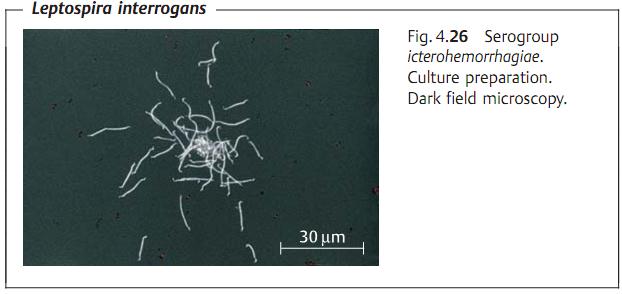
Morphology and culture. Leptospirae are fine spirochetes, 10-20 µm long, and 0.1-0.2 µm thick (Fig. 4.26). They possess no flagella, but rather derive their motility from rotating motions of the cell corpus. Visualization of lep-tospirae is best done using dark field or phase contrast microscopy. Lepto-spirae can be grown in special culture mediums under aerobic conditions at temperatures between 27-30 °C
Pathogenesis and clinical picture. Leptospirae invade the human organism through microinjuries in the skin or the intact conjunctival mucosa. There are no signs of inflammation in evidence at the portal of entry. The organisms spread to all parts of the body, including the central nervous system, hematogenously. Leptospirosis is actually a generalized vasculitis. The pathogens damage
mainly the endothelial cells of the capillaries, leading to greater permeability and hemorrhage and interrupting the O2 supply to the tissues. Jaundice is caused by a non-necrotic hepatocellular dysfunction. Disturbances of renal function result from hypoxic tubular damage. A clinical distinction is drawn between anicteric leptospirosis, which has a milder course, and the severe clinical picture of icteric leptospirosis (Weil disease). In principle, any of the serovars could potentially cause either of these two clinical courses. In practice, however, the sero-group icterohemorrhagiae is isolated more frequently in Weil disease.
Both types of leptospirosis are characterized by fever with chills, head-ache, and myalgias that set in after an incubation period of seven to 12 days. This initial septic stage of the disease lasts three to seven days and is then followed by the second, so-called immune stage, which lasts from four to 30 days. The most important clinical manifestation of stage two anicteric leptospirosis is a mild, aseptic meningitis. The second stage of Weil disease is characterized by hepatic and renal dysfunctions, extensive hemorrhaging, cardiovascular symptoms, and clouding of the state of consciousness. The immunity conferred by survival of the infection is reliable, but only protects against the one specific serovar.
Diagnosis. Detection and identification of leptospirae are accomplished by growing the organisms in cultures. Blood, cerebrospinal fluid, urine, or organ biopsies, which must not be contaminated with other bacteria, are incubated in special mediums at 27-30 °C for three to four weeks. A microscope check (dark field) is carried out every week to see if any leptospirae are proliferating. The Leptospirae are typed serologically in a lysis-agglutination reaction with specific test sera.
The method of choice for a laboratory diagnosis is an antibody assay. The antibodies produced after the first week of the infection are detected in patient serum using a quantitative lysis-agglutination test. Viable culture strains of the regionally endemic serovars provide the test antigens. The reaction is read off under the microscope.
Therapy. The agent of choice is penicillin G.
Epidemiology and prevention. Leptospiroses are typical zoonotic infections. They are reported from every continent in both humans and animals. The most important sources of infection are rodents and domestic animals, mainly pigs. The animals excrete the pathogen with urine. Leptospirae show little resistance to drying out so that infections only occur because of contact with a moist milieu contaminated with urine. The persons most at risk are farmers, butchers, sewage treatment workers, and zoo staff.
Prevention of these infections involves mainly avoiding contact with material containing the pathogens, control of Muridae rodents and successful treatment of domestic livestock. It is not necessary to isolate infected persons or their contacts. There is no commercially available vaccine.


|
|
|
|
كيف تعزز نمو الشعر الصحي؟
|
|
|
|
|
|
|
10 فحوصات مهمة يجب القيام بها لسيارتك قبل الصيف
|
|
|
|
|
|
العتبة العباسية تختتم فعاليات حفل التكليف الشرعي في قضاء عين التمر بكربلاء
|
|
|
|
طالبات مدارس عين التمر يرددن نشيد التكليف الشرعي
|
|
|
|
الطالبات المشاركات في حفل التكليف الشرعي يقدمن الشكر للعتبة العباسية
|
|
|
|
حفل التكليف الشرعي للطالبات يشهد عرض فيلم تعريفي بمشروع (الورود الفاطمية)
|