آخر المواضيع المضافة

 النبات
الحيوان
الأحياء المجهرية
علم الأمراض
التقانة الإحيائية
التقنية الحيوية المكروبية
التقنية الحياتية النانوية
علم الأجنة
الأحياء الجزيئي
علم وظائف الأعضاء
الغدد
المضادات الحيوية
النبات
الحيوان
الأحياء المجهرية
علم الأمراض
التقانة الإحيائية
التقنية الحيوية المكروبية
التقنية الحياتية النانوية
علم الأجنة
الأحياء الجزيئي
علم وظائف الأعضاء
الغدد
المضادات الحيوية| Calcium-Binding Proteins |


|
|
Read More
Date: 7-12-2015
Date: 16-12-2015
Date: 31-10-2020
|
Calcium-Binding Proteins
Calcium-binding proteins (CaBPs) are a key component linking the inorganic and organic systems that produce biological activities. Calcium is one of the most abundant inorganic elements in nature and is ubiquitous throughout biology, playing roles at the organismal, cellular, and molecular levels. Calcium is an essential component of shells and bones, where it is essentially deposited in crystals to create macroscopic support structures. At the cellular level, Ca2+ is one of the crucial currencies of most living organisms, acting as a second messenger in a wide range of key intracellular and extracellular systems. Calcium-binding proteins play important roles in mediating each of these effects. These proteins can be grouped into four primary categories: (1) intracellular proteins involved in Ca2+-mediated signal transduction, (2) enzymes, (3) extracellular cell surface and extracellular matrix proteins, and (4) proteins containing g-carboxyglutamic acid (Gla) residues. Although a variety of structural motifs bind Ca2+ , all involve oxygen atoms of the protein backbone or side chains, reflecting the intrinsic affinity of Ca2+ for oxygen atoms.
1. Intracellular Calcium-Signaling Proteins
1.1. EF-Hand Calcium-Binding Proteins
This family is the most extensively studied class of CaBPs. These proteins are characterized by a highly conserved helix–loop–helix motif termed the EF-Hand Motif, which consists of a 12-residue Ca2+-binding loop flanked by two alpha-helices. Almost all EF-hand CaBPs are composed of pairs of EF-hands. This pairing of Ca2+-binding sites is presumed to stabilize the protein conformation, increase the Ca2+ affinity of each site over that of isolated sites, and provide a means for cooperativity in Ca2+ binding. The cooperativity between sites allows for an “all or nothing” response to Ca2+-binding, which is crucial for the function of these CaBPs as intracellular Ca2+ sensors.
Calmodulin and troponin C are the best known members of this family of CaBPs. They each have two largely independent domains connected by a flexible linker. Each domain contains two EF-hands, so these proteins each bind four Ca2+ ions. In the resting cell, they exist in an inactive state, with either Mg2+ or no ion bound. When the intracellular Ca2+ concentration rises in response to a signal, the proteins bind Ca2+. This induces a dramatic conformational change, exposing a large hydrophobic surface within each domain that can interact with target proteins. There are many other CaBPs thought to function in a similar manner including caltractin and the calmodulin-like domain of plant Ca2+-dependent protein kinase.
Less is known about the S100 proteins, another large and important subfamily of EF-hand CaBPs. The proteins in this subfamily are composed of two EF-hands each, the first of which is a variant version with a 14-residue binding loop, termed a pseudo-EF-hand. The ligands in the pseudo EF-hand are mainly carbonyl oxygens of the peptide backbone, whereas the canonical 12-residue loop coordinates Ca2+ primarily with oxygen atoms of side chains. Many S100 proteins are found as hetero- or homodimers, and they exhibit tissue-specific expression patterns. While these proteins are thought to be involved in signal transduction, the molecular mechanism of this function is not known, nor have any target proteins been positively identified. However, the expression of S100 proteins is deregulated in some diseases, including cancer, rheumatoid arthritis, and Down's syndrome, and antibodies against these proteins are commonly used as markers for screening for these diseases.
Most EF-hand CaBPs are directly involved in intracellular calcium signal transduction. However, at least three fulfill other cellular requirements. Parvalbumin and calbindin D9k (an S100 protein) are thought to play roles in Ca2+ buffering and in Ca2+ uptake and transport, respectively. These and other members of the EF-hand protein family are active in various aspects of intracellular Ca2+ homeostasis. The diversity of the roles of EF-hand CaBPs is further illustrated by the recent structure of BM-40, a glycoprotein found in the extracellular matrix (1), which was shown to contain two EF-hands. Prior to this discovery, the EF-hand motif was thought to be unique to intracellular CaBPs. This diversity in function of the EF-hand CaBPs provides evidence of very extensive evolutionary optimization of the fit between the EF-hand CaBP fold and the calcium ion.
1.2. Annexins
The annexin family is a second important class of intracellular CaBPs. The exact function of the proteins in this family is unknown, but they do exhibit an intriguing Ca2+-dependent high-affinity binding to phospholipids. Annexins bind a large number of Ca2+ ions with high cooperativity. They assist transport of Ca2+ ions across membranes in vitro, although the physiological relevance has not yet been established. The mechanism of membrane binding is not known. The current model involves the Ca2+ ions acting as “glue” by simultaneously interacting with the protein and the membrane (2).
2. Ca2+-Binding Enzymes
Two well-known enzymes exhibit Ca2+-dependent translocation to the membrane fraction, presumably through a mechanism similar to that used by the annexins. Some isoforms of protein kinase C bind to phospholipids with high affinity in the presence of Ca2+ (2). The intracellular group IV phospholipase A2 also requires Ca2+ for membrane association (3).
Many other enzymes require Ca2+ ions for stability. This includes many serine proteases: members of both the trypsin and the subtilisin families have been found to bind Ca2+. These proteases each bind one to three Ca2+ ions, which are required for structural stability but do not directly participate in catalysis. In most cases, the Ca2+ ions are coordinated by ligands dispersed throughout the structure. Trypsin is an exception, however, and binds Ca2+ using a single 12-residue surface loop. The use of Ca2+ ions to stabilize the protein fold is not unique to the serine proteases. Another example of an enzyme that uses Ca2+ in this manner is thermolysin, a Zn2+-dependent protease. In other enzymes, the Ca2+ ions are directly involved in catalysis. This includes the secreted forms of phospholipase A2, in which the required Ca2+ ion is thought to be involved in transition state stabilization (4). Staphylococcal nuclease, a secreted protein that catalyzes DNA and RNA hydrolysis, also relies on Ca2+ for catalysis. The structure of this protein shows Ca2+ bound in the active site, where it is thought to be used to polarize the phosphate at the scissile phosphoester bond (5, 6).
3. Extracellular Cell Surface and Extracellular Matrix Ca2+-binding Proteins
3.1. Cadherins
These are tissue-specific cell adhesion molecules that are strongly Ca2+-dependent. At the levels of Ca2+ found in the extracellular milieu, cadherins bind several Ca2+ ions. A major conformational change is seen when Ca2+ is removed from these proteins. The resulting change to the apo state conformation is thought to prevent cadherins from interacting with each other.
3.2. C-Type Lectins
The C-type lectins are a Ca2+-dependent class of lectins, which mediate many cell surface carbohydrate recognition events. Selectins, concanavalin A, and mammalian mannose-binding protein are examples of this type of CaBP. The structure of mannose-binding protein shows that Ca2+ is directly involved in binding the carbohydrate to this protein. In concanavalin A, on the other hand, the Ca2+-binding site is 1.0–1.4 × 10⁻⁹ m from the carbohydrate binding site; Ca2+ does not participate directly in binding the carbohydrate, but instead stabilizes the protein structure. It is not known whether selectins use Ca2+ directly in carbohydrate binding or to stabilize the fold required for carbohydrate recognition.
3.3. EGF Modules
A subset of EGF motifs present as domains in a number of proteins contain b-hydroxy–aspartic acid residues and have Ca2+-binding activity. This type of EGF motif is found in some proteins involved in the blood clotting cascade and also in the extracellular matrix protein fibrillin. Fibrillin has 54 EGF modules, 43 of which are thought to bind Ca2+. It is thought that Ca2+ is important in maintaining the structure of the fibrillin monomers and also in allowing the monomers to aggregate into microfibrils. A sheath of these microfibrils covers and stabilizes the elastin fibers that give tissues such as skin, blood vessels, and lungs their needed elasticity.
4. Gla-Containing Proteins
The Gla-containing proteins contain g-carboxyglutamic acid residues. This is a glutamic acid residue that has been carboxylated in a vitamin K–dependent process. These proteins are found in the bones and teeth, in the kidney, and in the blood. Osteocalcin (bone Gla-protein) and matrix Gla-protein are found in bone. They are believed to be involved in the mineralization of this tissue (7). Similar proteins are thought to be involved in the mineralization of teeth. In the kidney, the Gla-containing protein nephrocalcin inhibits the nucleation, aggregation, and growth of calcium oxalate crystals. An abnormal form of this protein is found in the urine of people suffering from kidney stones. However, it is not entirely clear whether this defect is responsible for the growth of kidney stones (8).
By far the best studied of the Gla-containing proteins are those found in blood. The majority of Gla-containing proteins are zymogen forms of serine proteases involved in the blood clotting (coagulation) cascade. These proteins are: prothrombin, factor VII, factor IX, factor X, protein C, protein S, and protein Z. The protease domains of these proteins are very similar to the pancreatic digestive proteases trypsin, chymotrypsin, and elastase. The Gla-containing coagulants require Ca2+ binding in order to bind to phospholipids. This Ca2+-dependent phospholipid binding is responsible for the membrane association properties required for the function of the proteins. Ca2+ binding also appears to be necessary for the formation of the native conformation of the Gla domain (9) .
The Gla-containing coagulants have two classes of metal ion binding sites. There are approximately three higher affinity sites that are not metal ion–specific, and three to four lower affinity sites, which are specific for Ca2+. Only Sr2+ can substitute for Ca2+ in both types of binding site.
The high resolution three-dimensional structure of the Gla domain of prothrombin, with seven Ca2+ ions bound (10), reveals the Gla domain to involve nine to 10 turns of alpha-helix, in three separate helices. Seven Ca2+ ions interact with 24 oxygen atoms from 16 of the 18 carboxylate groups of the nine Gla residues that are ordered in the structure. A 10th Gla residue is disordered and does not participate in Ca2+ binding. The coordination geometries of the Ca2+ ions do not correspond to any idealized polyhedra. Five of the Ca2+ ions are involved in a polymeric array with 18 of the liganding oxygen atoms. Four of these Ca2+ ions are completely buried in the protein. This complex structure is thought to nucleate the folding of the Gla domain, and is essentially electrically neutral. The complexity and irregularity of this structure explains the selectivity for Ca2+ ions. Ca2+ is able to adopt different and distorted coordination geometries. Mg2+, on the other hand, is fairly rigid in its requirement for six ligands, and cannot accommodate the unusual network of ligands in the Gla domain. The remaining two metal ion sites in the Gla domain of prothrombin are solvent accessible, and carry a net charge of about +0.5 each. Because of the positive charge, these sites are thought to be involved in neutralizing the negatively-charged phospholipids, allowing the protein to associate with membranes.
References
1.E. Hohenester, P. Maurer, C. Hohenadl, R. Timpl, J. N. Jansonius, and J. Engel (1996) Nature Struct. Biol. 3, 67–73.
2.M. D. Bazzi and G. L. Nelsestuen (1993) Cell. Signal. 5, 357–365.
3.J. Y. Channon and C. C. Leslie (1990) J. Biol. Chem. 265, 5409–5413.
4.E. A. Dennis (1994) J. Biol. Chem. 269, 13057–13060.
5.P. J. Loll and E. E. Lattmam (1989) Prot. Struct. Funct. Genet. 5, 183–201.
6.F. A. Cotton, E. E. Hazen, and M. J. Legg (1979) Proc. Natl. Acad. Sci. USA 76, 2551–2555.
7. M. F. Young, J. M. Kerr, K. Ibaraki, A.-M. Heegaard, and P. G. Robey (1992) Clin. Orthoped. Rel. Res. 281, 275–294.
8. F. L. Coe, Y. Nakagawa, J. Asplin, and J. H. Parks (1994) Miner. Electrolyte Metab. 20, 378–384.
9. J. W. Suttie (1993) FASEB J. 7, 445–452.
10. M. Soriano-Garcia, K. Padmanabhan, A. M. de Vos, and A. Tulinsky (1992) Biochemistry 31, 2554–2566.


|
|
|
|
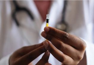
إجراء أول اختبار لدواء "ثوري" يتصدى لعدة أنواع من السرطان
|
|
|
|
|
|
|
دراسة تكشف "سببا غريبا" يعيق نمو الطيور
|
|
|
|
|
|
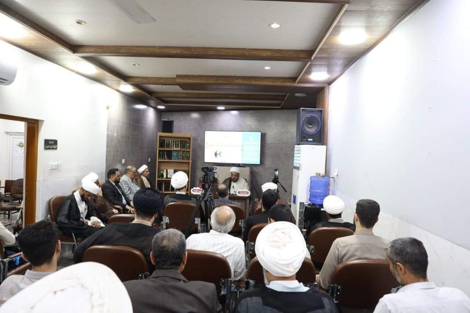
قسم الشؤون الفكرية يقيم برنامج (صنّاع المحتوى الهادف) لوفدٍ من محافظة ذي قار
|
|
|
|
الهيأة العليا لإحياء التراث تنظّم ورشة عن تحقيق المخطوطات الناقصة
|
|
|
|
قسم شؤون المعارف يقيم ندوة علمية حول دور الجنوب في حركة الجهاد ضد الإنكليز
|
|
|
|
وفد جامعة الكفيل يزور دار المسنين في النجف الأشرف
|