

 النبات
 الحيوان
 الأحياء المجهرية
 علم الأمراض
 التقانة الإحيائية
 التقنية الحيوية المكروبية
 التقنية الحياتية النانوية
 علم الأجنة
 الأحياء الجزيئي
 علم وظائف الأعضاء
 الغدد
الغدد
 المضادات الحيوية
|
Read More
Date: 2-5-2016
Date: 31-5-2021
Date: 22-12-2015
|
Beta-Bulge
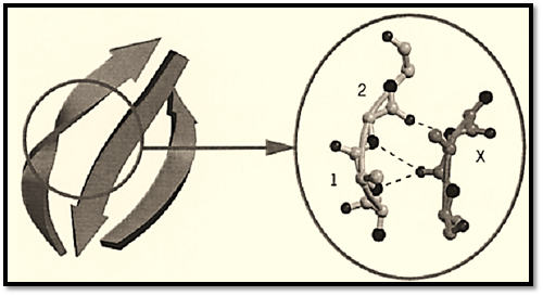
The b-bulge is a term used to define a specific secondary structure feature of protein structures, where the regular hydrogen bond interactions and backbone conformation of the b-sheet are disrupted by the presence of an extra residue. The additional residue is usually located on a b-strand at one edge of the b-sheet, where the bulge is more easily accommodated within the structure of the protein (Fig. 1). b-Bulges occur frequently, and often there are two or more per protein (1). In some cases, b-bulges have been found to be conserved in the structures of related proteins, where they may play a functional role. The more general role of the b-bulge is to alter the direction of the b-strand, so that it may be considered a type of turn. However, the change in direction of the polypeptide chain is not as pronounced as that caused by other types of turns.

Figure 1. Example of a classic b-bulge in an antiparallel b-sheet. Three b-strands of the antiparallel b-sheet are shown. The normal twist of the b-sheet is clearly accentuated by the b-bulge, caused by an additional residue in the middle b-strand at the edge of the b-sheet. The close-up view of the b-bulge (right) shows residue X from the b-strand forming backbone hydrogen bonds (shown as dashed lines) with two residues (1 and 2) from the outer b-strand. For clarity, side-chain atoms have been removed, backbone nitrogen atoms and backbone oxygen atoms are shown as dark spheres. This figure was generated using Molscript (3) and Raster3D (4, 5).
A b-bulge is that region between two consecutive b-type hydrogen bonds that includes two residues (called positions 1 and 2) on one b-strand opposite a single residue (position X) on the other b-strand (2). There are several different classes of b-bulge, most (90%) occurring between antiparallel b-strands. The two most common are the classic and the G1, accounting for about 80% of b-bulges. In the classic b-bulge, the residue at position 1 has backbone dihedral angles (f = –100, y = –25), closer to those of an a-helical than to a b-strand conformation, but residue 2 has angles closer to the b-strand conformation (f = –180 and y = 160). In the G1 b-bulge, the residue at position 1 has a positive f value (f = 85 and y = 0) and is therefore almost always glycine (thus the name G1). The residue at position 2 of the G1 class has dihedral angles corresponding to b-strand (y = –90 and y = 150). The G1 b-bulge often occurs in combination with a type II b-turn (which requires a glycine at position 3). Compared to the usual b-sheet structure, a b-bulge disrupts the alternating side chain placement on one of the b-strands and increases the right-handed twist of the b-strand from the usual 10° to 35° to 45°.
References
1.A. W. E. Chan, E. G. Hutchinson, D. Harris, and J. M. Thornton (1993) Protein Sci. 2, 1575–1590.
2.J. S. Richardson, E. D. Getzoff, and D. C. Richardson (1978) Proc. Natl. Acad. Sci. USA 75, 2574-2578.
3. P. J. Kraulis (1991) J. Appl. Crystallogr. 24, 946–950.


|
|
|
|
التوتر والسرطان.. علماء يحذرون من "صلة خطيرة"
|
|
|
|
|
|
|
مرآة السيارة: مدى دقة عكسها للصورة الصحيحة
|
|
|
|
|
|
|
نحو شراكة وطنية متكاملة.. الأمين العام للعتبة الحسينية يبحث مع وكيل وزارة الخارجية آفاق التعاون المؤسسي
|
|
|