

 النبات
 الحيوان
 الأحياء المجهرية
 علم الأمراض
 التقانة الإحيائية
 التقنية الحيوية المكروبية
 التقنية الحياتية النانوية
 علم الأجنة
 الأحياء الجزيئي
 علم وظائف الأعضاء
 الغدد
الغدد
 المضادات الحيوية
|
Read More
Date: 2025-02-08
Date: 2025-01-25
Date: 2025-04-08
|
Definition
• A malignant epithelial tumour arising in the lung.
Epidemiology
• One of the most common and deadly cancers with nearly 2 million deaths annually. • Most present in patients aged over 60 years.
Aetiology
• About 80% of cases are directly attributable to smoking.
Classification
• Adenocarcinoma (40%).
• Squamous cell carcinoma (20%).
• Small cell carcinoma (15%).
• A number of rare subtypes make up the remainder.
Carcinogenesis
• Similar to other carcinomas, lung carcinomas are likely to arise from a precursor phase of epithelial dysplasia, representing neoplastic transformation of lung epithelium without invasion.
• Adenomatous dysplasia/adenocarcinoma in situ precedes adenocarcinoma.
• Squamous dysplasia/ squamous carcinoma in situ precedes squamous cell carcinoma.
Genetic mutations
• Adenocarcinoma: Gain of function mutations in receptor tyrosine kinase genes such as EGFR, ALK, ROS, and MET.
• Squamous cell carcinoma: Loss of function mutations in tumour suppressor genes such as TP53 and CDKN2A.
• Small cell carcinoma: Inactivation of TP53 and RB; amplification of MyC family.
Presentation
• Symptoms related to local growth of the tumour include progressive breathlessness, cough, chest pain, hoarseness, or loss of voice, haemoptysis, weight loss, and recurrent pneumonia.
• Abdominal pain, bony pain, and neurological symptoms may occur from metastases.
• A small proportion of small cell carcinomas present with paraneoplastic syndromes or the superior vena cava syndrome.
Macroscopy
• A firm white/ grey tumour mass within the lung.
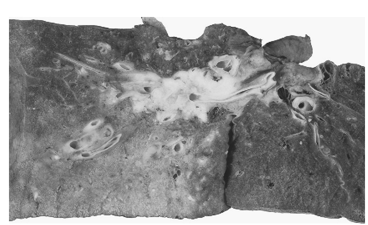
• yellow consolidation may be seen in the lung parenchyma distal to large proximal tumours due to an obstructive pneumonia (Fig.1).
• Pleural puckering may be seen overlying peripheral tumours that have infiltrated the pleura.
• Metastatic tumour deposits may be seen in hilar lymph nodes.

Fig.1 A central lung carcinoma. note how the lung tissue distal to the tumour shows flecks of yellow consolidation due to an obstructive pneumonia. the tumour was found to be a squamous cell carcinoma when examined microscopically. Reproduced with permission from Clinical Pathology (Oxford Core texts), Carton, James, Daly, Richard, and Ramani, Pramila, Oxford University Press (2006), p.131, Figure 7.13.
Histopathology
• Adenocarcinoma: malignant epithelial tumour showing glandular differentiation and/ or mucin production.
• Squamous cell carcinoma: malignant epithelial tumour showing keratinization and/ or intercellular bridges.
• Small cell carcinoma: high grade neuroendocrine carcinoma composed of small cells with scant cytoplasm, ill- defined cell borders, finely granular chromatin, and absent nucleoli. Mitotic activity is high and necrosis is often extensive.
Immunohistochemistry
• the main histological types of lung carcinomas show differing patterns of immunohistochemistry which can aid in diagnosis.
• Adenocarcinoma: p63/ p40 negative; TTF1 positive.
• Squamous cell carcinoma: p63/ p40 positive; TTF1 negative. • Small cell carcinoma: neuroendocrine marker (CD56, chromogranin, synaptophysin) positive; TTF1 positive.
Prognosis
• Poor with 5- year survival rates of ~10% in most countries.


|
|
|
|
التوتر والسرطان.. علماء يحذرون من "صلة خطيرة"
|
|
|
|
|
|
|
مرآة السيارة: مدى دقة عكسها للصورة الصحيحة
|
|
|
|
|
|
|
نحو شراكة وطنية متكاملة.. الأمين العام للعتبة الحسينية يبحث مع وكيل وزارة الخارجية آفاق التعاون المؤسسي
|
|
|