

 النبات
 الحيوان
 الأحياء المجهرية
 علم الأمراض
 التقانة الإحيائية
 التقنية الحيوية المكروبية
 التقنية الحياتية النانوية
 علم الأجنة
 الأحياء الجزيئي
 علم وظائف الأعضاء
 الغدد
الغدد
 المضادات الحيوية
|
Read More
Date: 17-11-2015
Date: 22-3-2016
Date: 22-3-2016
|
Subcutaneous mycoses are fungal infections of the dermis, subcutaneous tissue, and bone. Causative organisms reside in the soil and decaying or live vegetation.
A. Epidemiology
Subcutaneous fungal infections are almost always acquired through traumatic lacerations or puncture wounds. Sporotrichosis, for example, is often acquired from the prick of a thorn. As expected, these infections are more common in individuals who have frequent con tact with soil and vegetation and wear inadequate protective clothing. The subcutaneous mycoses are not transmissible from human to human.
B. Clinical significance
With the rare exception of sporotrichosis, which shows a broad geo graphic distribution in the United States, the common subcutaneous mycoses discussed below are confined to tropical and subtropical regions.
1. Sporotrichosis:
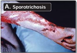
This infection, characterized by a granulomatous ulcer at the puncture site, may produce secondary lesions along the draining lymphatics (Figure 20.6A). The causative organism, Sporothrix schenckii, is a dimorphic fungus that exhibits the yeast form in infected tissue (see Figure 20.7) and the mycelial form upon laboratory culture. In most patients, the disease is self-limiting but may persist in a chronic form. Dissemination to distant sites is possible in patients with deficiencies in T-cell function (such as in AIDS and lymphomas). Oral itraconazole is the drug of choice.

2. Chromomycosis:
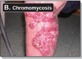
Also called chromoblastomycosis, this infection is characterized by warty nodules that spread slowly along the lymphatics and develop crusty abscesses (see Figure 20.6B). Pathogens causing this mycosis include several species of pigmented soil fungi (for example, Phialophora and Cladosporium), and the infection is most commonly seen in the tropics. Treatment is difficult. Surgical removal of small lesions is effective but must be performed cautiously and with wide margins to prevent dis semination. More advanced stages of the disease are treated with itraconazole and terbinafine.

3. Mycetoma (“Madura foot”):
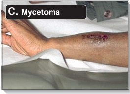
Mycetoma appears as a localized abscess, usually on the feet, but is not limited to the lower extremity (see Figure 20.6C). The abscess discharges pus, serum, and blood through sinuses (in this usage, sinus means “abnormal channel”). The infection can spread to the underlying bone and results in crippling deformities . The pathogenic agents are various soil fungi. Most common are Madurella grisea and Exophiala jeanselmei. Mycetomas appear similar to the lesions of chromomycosis, but the defining characteristic of mycetoma is the presence of colored grains, composed of compacted hyphae, in the exudate. The color of the grains (black, white, red, or yellow) is characteristic of the causative organism and, therefore, useful in identifying the particular pathogen. There is no effective chemotherapy for fungal mycetoma. Treatment is usually surgical excision.


|
|
|
|
التوتر والسرطان.. علماء يحذرون من "صلة خطيرة"
|
|
|
|
|
|
|
مرآة السيارة: مدى دقة عكسها للصورة الصحيحة
|
|
|
|
|
|
|
نحو شراكة وطنية متكاملة.. الأمين العام للعتبة الحسينية يبحث مع وكيل وزارة الخارجية آفاق التعاون المؤسسي
|
|
|