

 النبات
 الحيوان
 الأحياء المجهرية
 علم الأمراض
 التقانة الإحيائية
 التقنية الحيوية المكروبية
 التقنية الحياتية النانوية
 علم الأجنة
 الأحياء الجزيئي
 علم وظائف الأعضاء
 الغدد
الغدد
 المضادات الحيوية
|
Read More
Date: 9-10-2015
Date: 11-10-2015
Date: 29-1-2021
|
Cell Junctions
Cell junctions can be divided into two types: those that link cells together, also called intercellular junctions (tight, gap, adherens, and desmosomal junctions), and those that link cells to the extracellular matrix (focal contacts/adhesion plaques and hemidesmosomes). These junctions play a prominent role in maintaining the integrity of tissues in multicellular organisms and some, if not all of them, are involved in signal transduction.
Intercellular junctions and hemidesmosomes were first identified in tissues examined by electron microscopy. In contrast, the focal contact was first observed in cultured cells in the light microscope by a technique called interference reflection. This procedure revealed specific sites where cells closely adhere to their substrate. These were called focal contacts or adhesion plaques.
Tight Junctions
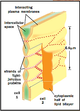
The tight junction (also referred to as a zonula occludens) is a site where the membranes of two cells come very close together. In fact, the outer leaflets of the membranes of the contacting cells appear to be fused. Tight junctions, as their name implies, act as a barrier so that materials cannot pass between two interacting cells. The protein components of the tight junction are arranged like beads on a string that span the adjacent membranes of each tight junction.
Tight junctions often occur in a belt completely encircling the cell. In a sheet of such cells, material cannot pass from one side of the sheet to the other by squeezing between cells. Instead, it must go through a cell, and hence the cell can regulate its passage. Such an arrangement is found in the gut, to regulate absorption of digested nutrients.

A model of a tight junction. It is thought that the strands that hold adjacent plasma membranes together are formed by continuous strands of transmembrane junctional proteins across the intercellular space, creating a seal.
Gap Junctions
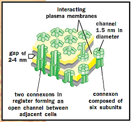
In contrast to the tight junction, there is a channel between the membranes of contacting cells in the gap junction so that the cytoplasm of the two is connected. The basic building block of each gap junction is the connexin subunit. Six of these in each of the membranes of two neighboring cells come together, and then the group of six connexins in one cell interacts with a comparable hexamer in the other cell resulting in the formation of a channel. This channel allows direct cytoplasmic communication among the cells; small molecules of 1,500 Daltons or less can pass through the channel of each gap junction whose opening or closing can be controlled locally in the cell. Gap junctions unite muscle cells in the heart to help coordinate their contraction.

Model of a gap junction. The lipid bilayers are penetrated by protein assemblies called connexons. Two connexons join across the intercellular space to form a continuous aqueous channel that links the two cells.
Adherens Junctions and Focal Contacts
Adherens junctions (sometimes called zonula adherens) are found at sites of cell-cell interaction. Focal contacts mediate association of cells with the extracellular matrix. Both associate with the actin cytoskeleton and both are involved in adhesion (sticking cells together or sticking cells to surfaces). Focal contacts possess specific transmembrane receptors of the integrin family that link the cell to the extracellular matrix on the outside of the cell and the microfilament system on the inside. Conversely, members of a family of calcium ion-dependent cell adhesion molecules, called cadherins, mediate attachment between cells at adherens junctions. Adherens junctions and focal contacts not only tether cells together or to the extracellular matrix, but they also transduce signals into and out of the cell, influencing a variety of cellular behaviors including proliferation, migration, and differentiation. In fact some protein components of these junctions can shuttle to and from the nucleus where they are thought to play a role in regulating gene expression.
Desmosomes and Hemidesmosomes
Desmosomes (the macula adherens) and hemidesmosomes are distinguished by their association with the keratin-based cytoskeleton. Despite their names, desmosomes and hemidesmosomes are distinct at the molecular level. Both are primarily involved in adhesion. The desmosome, like the adherens junction, possesses calcium ion-dependent cell adhesion molecules that interact with similar molecules in the adjacent cell. Meanwhile, integrins at the core of the hemidesmosome mediate its interaction with the extracellular matrix. The hemidesmosome and, most likely, the desmosome are also sites of signal transduction.
References
Alberts, Bruce, et al. Molecular Biology of the Cell, 4th ed. New York: Garland Publishing, 2000.


|
|
|
|
التوتر والسرطان.. علماء يحذرون من "صلة خطيرة"
|
|
|
|
|
|
|
مرآة السيارة: مدى دقة عكسها للصورة الصحيحة
|
|
|
|
|
|
|
نحو شراكة وطنية متكاملة.. الأمين العام للعتبة الحسينية يبحث مع وكيل وزارة الخارجية آفاق التعاون المؤسسي
|
|
|