آخر المواضيع المضافة

 النبات
الحيوان
الأحياء المجهرية
علم الأمراض
التقانة الإحيائية
التقنية الحيوية المكروبية
التقنية الحياتية النانوية
علم الأجنة
الأحياء الجزيئي
علم وظائف الأعضاء
الغدد
المضادات الحيوية
النبات
الحيوان
الأحياء المجهرية
علم الأمراض
التقانة الإحيائية
التقنية الحيوية المكروبية
التقنية الحياتية النانوية
علم الأجنة
الأحياء الجزيئي
علم وظائف الأعضاء
الغدد
المضادات الحيوية| Prokaryotic DNA Replication : Replication fork formation |


|
|
Read More
Date: 26-8-2021
Date: 10-11-2021
Date: 8-10-2021
|
Prokaryotic DNA Replication : Replication fork formation
As the two strands unwind and separate, synthesis occurs at two replication forks that move away from the origin in opposite directions (bidirectionally), generating a replication bubble . [Note: The term “replication fork” derives from the Y-shaped structure in which the tines of the fork represent the separated strands (Fig. 1).]

Figure 1: Proteins responsible for maintaining the separation of the parental strands and unwinding the double helix ahead of the advancing replication fork (). ADP = adenosine diphosphate; Pi = inorganic phosphate.
1. Required proteins: Initiation of DNA replication requires the recognition of the origin (start site) by a group of proteins that form the prepriming complex. These proteins are responsible for melting at the ori, maintaining the separation of the parental strands, and unwinding the double helix ahead of the advancing replication fork. In E. coli, these proteins include the following.
a. DnaA protein: DnaA protein initiates replication by binding to specific nucleotide sequences (DnaA boxes) within oriC. Binding causes an AT-rich region (the DNA unwinding element) in the origin to melt. Melting (strand separation) results in a short, localized region of ssDNA.
b. DNA helicases: These enzymes bind to ssDNA near the replication fork and then move into the neighboring double-stranded region, forcing the strands apart (in effect, unwinding the double helix). Helicases require energy provided by ATP hydrolysis (see Fig. 1). Unwinding at the replication fork causes supercoiling in other regions of the DNA molecule. [Note: DnaB is the principal helicase of replication in E. coli. Binding of this hexameric protein to DNA requires DnaC.]
c. Single-stranded DNA–binding protein: This protein binds to the ssDNA generated by helicases (see Fig. 1). Binding is cooperative (that is, the binding of one molecule of single-stranded binding [SSB] protein makes it easier for additional molecules of SSB protein to bind tightly to the DNA strand). The SSB proteins are not enzymes, but rather serve to shift the equilibrium between dsDNA and ssDNA in the direction of the single-stranded forms. These proteins not only keep the two strands of DNA separated in the area of the replication origin, thus providing the single-stranded template required by polymerases, but also protect the DNA from nucleases that degrade ssDNA.
2. Solving the problem of supercoils: As the two strands of the double helix are separated, a problem is encountered, namely, the appearance of positive supercoils in the region of DNA ahead of the replication fork as a result of overwinding (Fig. 2) and negative supercoils in the region behind the fork. The accumulating positive supercoils interfere with further unwinding of the double helix. [Note: Supercoiling can be demonstrated by tightly grasping one end of a helical telephone cord while twisting the other end. If the cord is twisted in the direction of tightening the coils, the cord will wrap around itself in space to form positive supercoils. If the cord is twisted in the direction of loosening the coils, the cord will wrap around itself in the opposite direction to form negative supercoils.] To solve this problem, there is a group of enzymes called DNA topoisomerases, which are responsible for removing supercoils in the helix by transiently cleaving one or both of the DNA strands.

Figure 2: Positive supercoiling resulting from DNA strand separation.
a. Type I DNA topoisomerases: These enzymes reversibly cleave one strand of the double helix. They have both strand-cutting and strandresealing activities. They do not require ATP, but rather appear to store the energy from the phosphodiester bond they cleave, reusing the energy to reseal the strand (Fig. 3). Each time a transient nick is created in one DNA strand, the intact DNA strand is passed through the break before it is resealed, thus relieving (relaxing) accumulated supercoils. Type I topoisomerases relax negative supercoils (that is, those that contain fewer turns of the helix than does relaxed DNA) in E. coli and both negative and positive supercoils (that is, those that contain fewer or more turns of the helix than does relaxed DNA) in many prokaryotic cells (but not E. coli) and in eukaryotic cells.

Figure 3: Action of type I DNA topoisomerases.
b. Type II DNA topoisomerases: These enzymes bind tightly to the DNA double helix and make transient breaks in both strands. The enzyme then causes a second stretch of the DNA double helix to pass through the break and, finally, reseals the break (Fig. 4). As a result, both negative and positive supercoils can be relieved by this ATP-requiring process. DNA gyrase, a type II topoisomerase found in bacteria and plants, has the unusual property of being able to introduce negative supercoils into circular DNA using energy from the hydrolysis of ATP. This facilitates the replication of DNA because the negative supercoils neutralize the positive supercoils introduced during opening of the double helix. It also aids in the transient strand separation required during transcription .

Figure 4: Action of type II DNA topoisomerase.
Anticancer agents, such as the camptothecins, target human type I topoisomerases, whereas etoposide targets human type II topoisomerases. Bacterial DNA gyrase is a unique target of a group of antimicrobial agents called fluoroquinolones (for example, ciprofloxacin).


|
|
|
|
دراسة: عدم ترتيب الغرفة قد يدل على مشاكل نفسية
|
|
|
|
|
|
|
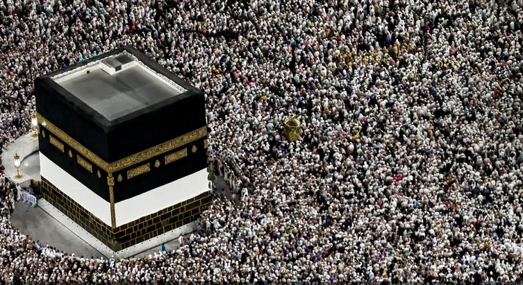
علماء: تغير المناخ تسبب في ارتفاع الحرارة خلال موسم الحج
|
|
|
|
|
|
|
جهود مكثفة وطباعة عشرات الآلاف من المنشورات .. استعدادات العتبة العلوية المقدسة لعيد الغدير الأغر
|
|
|