

النبات

مواضيع عامة في علم النبات

الجذور - السيقان - الأوراق

النباتات الوعائية واللاوعائية

البذور (مغطاة البذور - عاريات البذور)

الطحالب

النباتات الطبية


الحيوان

مواضيع عامة في علم الحيوان

علم التشريح

التنوع الإحيائي

البايلوجيا الخلوية


الأحياء المجهرية

البكتيريا

الفطريات

الطفيليات

الفايروسات


علم الأمراض

الاورام

الامراض الوراثية

الامراض المناعية

الامراض المدارية

اضطرابات الدورة الدموية

مواضيع عامة في علم الامراض

الحشرات


التقانة الإحيائية

مواضيع عامة في التقانة الإحيائية


التقنية الحيوية المكروبية

التقنية الحيوية والميكروبات

الفعاليات الحيوية

وراثة الاحياء المجهرية

تصنيف الاحياء المجهرية

الاحياء المجهرية في الطبيعة

أيض الاجهاد

التقنية الحيوية والبيئة

التقنية الحيوية والطب

التقنية الحيوية والزراعة

التقنية الحيوية والصناعة

التقنية الحيوية والطاقة

البحار والطحالب الصغيرة

عزل البروتين

هندسة الجينات


التقنية الحياتية النانوية

مفاهيم التقنية الحيوية النانوية

التراكيب النانوية والمجاهر المستخدمة في رؤيتها

تصنيع وتخليق المواد النانوية

تطبيقات التقنية النانوية والحيوية النانوية

الرقائق والمتحسسات الحيوية

المصفوفات المجهرية وحاسوب الدنا

اللقاحات

البيئة والتلوث


علم الأجنة

اعضاء التكاثر وتشكل الاعراس

الاخصاب

التشطر

العصيبة وتشكل الجسيدات

تشكل اللواحق الجنينية

تكون المعيدة وظهور الطبقات الجنينية

مقدمة لعلم الاجنة


الأحياء الجزيئي

مواضيع عامة في الاحياء الجزيئي


علم وظائف الأعضاء


الغدد

مواضيع عامة في الغدد

الغدد الصم و هرموناتها

الجسم تحت السريري

الغدة النخامية

الغدة الكظرية

الغدة التناسلية

الغدة الدرقية والجار الدرقية

الغدة البنكرياسية

الغدة الصنوبرية

مواضيع عامة في علم وظائف الاعضاء

الخلية الحيوانية

الجهاز العصبي

أعضاء الحس

الجهاز العضلي

السوائل الجسمية

الجهاز الدوري والليمف

الجهاز التنفسي

الجهاز الهضمي

الجهاز البولي


المضادات الميكروبية

مواضيع عامة في المضادات الميكروبية

مضادات البكتيريا

مضادات الفطريات

مضادات الطفيليات

مضادات الفايروسات

علم الخلية

الوراثة

الأحياء العامة

المناعة

التحليلات المرضية

الكيمياء الحيوية

مواضيع متنوعة أخرى

الانزيمات
Abdominal Vasculature and Lymphatics
المؤلف:
Kelly M. Harrell and Ronald Dudek
المصدر:
Lippincott Illustrated Reviews: Anatomy
الجزء والصفحة:
15-7-2021
5879
Abdominal Vasculature and Lymphatics
Arterial blood supply to abdominal viscera originates from the abdominal aorta. The thoracic aorta enters the abdomen through the aortic hiatus in the diaphragm at the T12 vertebral level. At this point, it is designated as the abdominal aorta, which extends inferiorly until bifurcating into right and left common iliac arteries at the L4 vertebral level. The abdominal aorta continues inferiorly at the bifurcation in the form of a small, often wanting, median sacral artery. Along the length of the abdominal aorta arise three anterior unpaired branches and multiple paired branches.
Venous drainage of structures in the abdominal cavity is unique in that it involves two systems-the portal system and the caval system.
A. Unpaired aortic vessels
Three unpaired branches arise from the anterior surface of the aorta and serve as the primary blood supply to organs associated with the foregut, midgut, and hindgut (Fig. 1). To ensure collateral blood flow, select viscera receive branches from more than one of these unpaired branches.
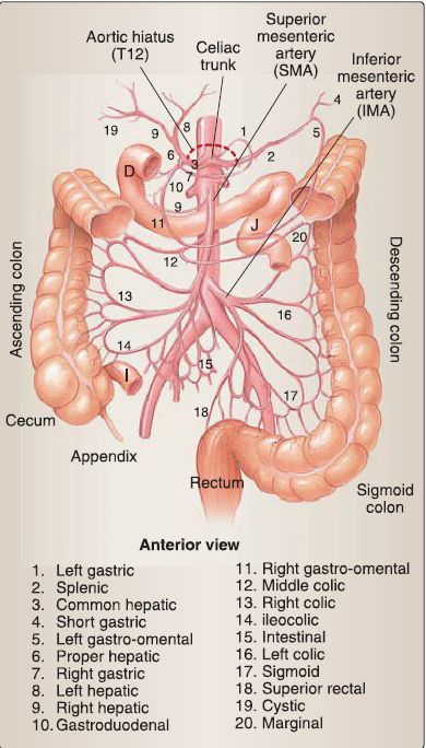
Figure 1: Abdominal arterial supply overview. D = duodenum, I = ileum, J = jejunum.
1. Celiac trunk: Short in length, the celiac trunk serves as the primary artery to foregut structures (Fig. 2). It is the first of the three unpaired vessels to branch from the abdominal aorta, just inferior to the aortic hiatus (T12 level). It almost immediately branches into the left gastric, splenic, and common hepatic arteries.
a. Left gastric artery: The left gastric artery courses to the left half of the lesser curvature of the stomach and gives off a small esophageal branch proximally. It anastomoses along the lesser curvature with the right gastric artery.
b. Splenic artery: This tortuous artery travels to the left, posteriorly to the pancreas and enters the splenorenal ligament before terminating in the hilum of the spleen. Just before entering the spleen, the artery gives off short gastric branches superiorly to the fundus of the stomach and a left gastro-omental (gastroepiploic) artery to the left greater curvature of the stomach.
This will eventually anastomose with the right gastro-omental (gastroepiploic) artery. Additional branches include dorsal and greater pancreatic arteries.
c. Common hepatic artery: This artery courses to the right, toward the liver. It divides into proper hepatic artery and gastroduodenal artery. Proper hepatic ascends toward the liver and gives off a right gastric artery before dividing into right and left hepatic arteries. Right hepatic artery typically gives rise to the cystic artery, which supplies the gallbladder.
Gastroduodenal gives off supraduodenal branches, descends between the duodenum (first part) and pancreas, and divides into superior pancreaticoduodenal and right gastro-omental (gastroepiploic) arteries.

Figure 2 : Celiac trunk (CT). CBD = common bile duct, CD = cystic duct, CH = common hepatic artery, CHD = common hepatic duct, GB = gallbladder, LG = left gastric, PH = proper hepatic artery, SA = splenic artery. * = variation of left hepatic artery, which typically is a branch of PH.
2. Superior mesenteric artery: The superior mesenteric artery (SMA) arises from the aorta, just inferior to the celiac trunk (L 1 level), and serves as the primary artery to midgut structures (Fig. 3). The artery travels inferiorly and crosses anterior to the left renal vein, pancreas (uncinate process), and the duodenum (third part, horizontal). Within the mesentery of the small intestine, the SMA gives off multiple branches.

Figure 3: Superior mesenteric artery. D = duodenum, P = pancreas.
a. Inferior pancreaticoduodenal: The first branch of the SMA travels superiorly to supply the duodenum and head/uncinate process of the pancreas. It anastomoses with the superior pancreaticoduodenal artery. This is an example of structures receiving blood supply from more than one unpaired aortic arteries.
b. Intestinal branches: Multiple (15-18) arteries course in the mesentery of the small intestine to supply the jejunum and ileum. Close to the organ, these vessels form a network of arterial arcades that extend into straight vessels called vasa recta. The pattern of arcades and vasa recta differs in the jejunum and ileum.
c. lleocolic artery: Found descending into the right lower quadrant, this artery serves as the primary supply to the distal ileum, cecum, and appendix. At the ileocecal junction, it gives off a small appendicular artery, which courses through the mesoappendix.
d. Right colic artery: This artery can be a direct branch of the SMA or arise from the ileocolic artery. It travels to supply the ascending colon (large intestines).
e. Middle colic artery: This artery arises from the proximal SMA and travels in the transverse mesocolon to supply the proximal two thirds of the transverse colon.
3. Inferior mesenteric artery: The inferior mesenteric artery arises from the aorta at the L3 level, just inferior to the duodenum (third part), as shown in Figure 4. It travels toward the left lower quadrant to supply hindgut structures. Its branches are as follows:
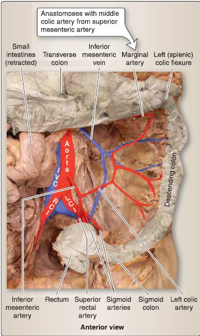
Figure 4 : Inferior mesenteric artery. IVG = inferior vena cava, LGI = left common iliac artery, RGI = right common iliac artery.
a. Left colic artery: This artery supplies the distal third of the transverse colon (by way of the marginal artery) and descending colon.
b. Sigmoid arteries: This collection of arteries supplies the sigmoid colon.
c. Superior rectal artery: This artery supplies the superior rectum and anastomoses with middle and inferior rectal arteries to provide collateral blood flow to the rectum.
B. Paired aortic vessels
As shown in Figure 5, paired vessels from the aorta supply the abdominal body wall and paired viscera in the abdominal cavity, such as the kidneys, or viscera that originated in the abdominal cavity during development, such as the gonads (ovaries/testes). The following paired vessels are listed from superior to inferior along the length of the abdominal aorta.
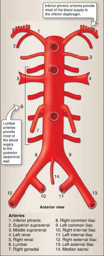
Figure 5: Paired aortic branches.
1. Inferior phrenic arteries: These supply the diaphragm and gives off superior suprarenal arteries.
2. Middle suprarenal arteries: These arise at the L 1 level to supply the suprarenal glands.
3. Renal arteries: These large vessels arise at the L 1 level to supply the kidneys. The right renal artery travels posterior to the IVG to reach the right kidney. Renal arteries also give off inferior suprarenal arteries.
4. Lumbar arteries: From L 1 to L4 levels, four to five paired arteries arise from the aorta to supply the posterior abdominal wall and surrounding spinal structures.
5. Gonadal arteries: Ovarian (female) and testicular (male) arteries originate from the abdominal aorta at the L2 level, despite the final position of the ovaries in the pelvis and testes in the scrotum.
C. Portal-caval system
Two venous systems exist in the abdomen. The portal venous system is responsible for transporting venous blood from the GI viscera (including pancreas and spleen) to the liver for filtration by way of the hepatic portal vein. The caval venous system drains venous blood from structures of the posterior abdominal wall, kidneys, gonads, and suprarenal glands. Important anastomoses between these two systems occur at select sites in the abdomen and pelvis to ensure collateral flow in the event of blockage.
1. Portal venous system: The hepatic portal vein is most commonly formed by the union of the superior mesenteric and splenic veins (Fig. 6). The inferior mesenteric vein typically drains into either the splenic vein, superior mesenteric vein, or directly into the union of these vessels. Tributaries of these main vessels collect blood from abdominal viscera to be eventually filtered through the liver. The hepatic portal vein travels in the hepatoduodenal ligament to reach the liver, where it divides into right and left portal veins.
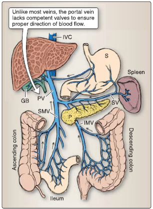
Figure 6: Portal venous system. Arrows indicate direction of venous blood flow from viscera to liver by way of the portal vein (PV). GB = gallbladder, IMV = inferior mesenteric vein, IVC = inferior vena cava, P = pancreas, S = stomach, SMV = superior mesenteric vein, SV = splenic vein.
2. Caval venous system: The caval venous system is made up of the IVC and its tributaries (Fig. 7). The IVC is formed by the union of the left and right common iliac veins. In addition to draining blood from the posterior body wall, kidneys, gonads, and suprarenal glands, it receives venous blood from pelvic and perinea! structures and the lower limbs. This venous blood bypasses the liver to enter the right atrium of the heart. In the abdomen, the IVC is situated to the right of the aorta, travels posterior to the liver, and receives multiple, short hepatic veins from the liver before piercing the diaphragm at the TS level to enter the heart.
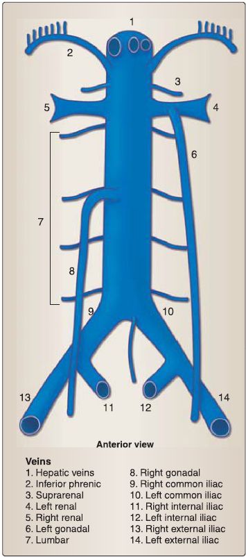
Figure 7: Caval venous system.
3. Portal-caval anastomoses: Venous anastomoses between these two systems occur in three main regions-the distal esophagus, paraumbilicus, and rectum (Fig. 8).
a. Esophagus anastomoses: Tributaries of the left gastric vein (portal system) anastomose with esophageal veins (caval system) of the azygos system.
b. Paraumbilical anastomoses: Paraumbilical veins (portal system) anastomose with superficial veins of the anterolateral abdominal wall (caval system).
c. Rectal anastomoses: Superior rectal veins (portal system) anastomose with middle and inferior rectal veins (caval system).

Figure 8: Portal-caval anastomoses. Stars indicate site of anastomosis.
D. Abdominal lymphatics
Lymph nodes are located throughout the abdomen to receive lymph from abdominal viscera and body wall structures (Fig. 9).
1. Preaortic nodes: Central collections of preaortic nodes include celiac, superior mesenteric, and inferior mesenteric nodes, which are located adjacent to the arteries of the same name.
2. Para-aortic nodes: Central para-aortic nodes include right (caval) and left (aortic) lumbar nodes, which are located along the length of the IVC and aorta, respectively.
3. Peripheral nodes: Peripheral collections of nodes are scattered throughout the mesenteries and along vessels, closer to viscera. These nodes drain centrally.
4. Cysterna chyli: Efferents from preaortic nodes (celiac, superior mesenteric, inferior mesenteric) join to form right and left intestinal lymph trunks. Efferents from lumbar nodes join to form right and left lumbar lymph trunks. These trunks coalesce at the abdominal confluence, the site of a small lymphatic sac called the cysterna chyli. Although commonly less sac-like and more plexiform in appearance, the cysterna chyli sits just inferior to the diaphragm at the aortic hiatus (T12 level). Here, it transitions into the thoracic duct, which continues superiorly in the thoracic cavity.
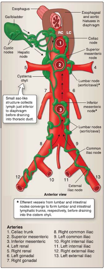
Figure 9: Lymphatics of abdominal viscera. Black arrows indicate general direction
of lymphatic flow. LC = left crus, RC = right crus.
 الاكثر قراءة في علم التشريح
 اخر الاخبار
اخبار العتبة العباسية المقدسة

الآخبار الصحية


 قسم الشؤون الفكرية يصدر كتاباً يوثق تاريخ السدانة في العتبة العباسية المقدسة
 "المهمة".. إصدار قصصي يوثّق القصص الفائزة في مسابقة فتوى الدفاع المقدسة للقصة القصيرة
 (نوافذ).. إصدار أدبي يوثق القصص الفائزة في مسابقة الإمام العسكري (عليه السلام)


