Cranium
The cranium, or skull, comprises a collection of singular and paired bones that articulate and fuse over the course of development. Covering the top part of the skull is the multilayered scalp.
A. Osteology
The skull comprises two parts: the neurocranium and the viscerocranium. Bones that contribute to the complete formation of the skull include (Fig. 1) paired (parietal, temporal, maxilla, inferior turbinate [nasal concha], zygomatic, palatine, nasal, and lacrimal) and singular (frontal, ethmoid, sphenoid, occipital, mandible, and vomer) bones.
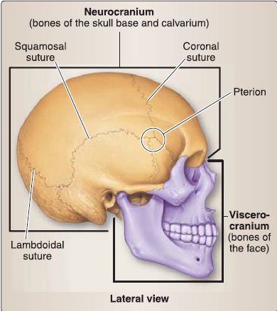
Figure 1: Adult cranium showing neurocranium and viscerocranium and associated sutures.
1. Embryology: The neurocranium consists of the flat bones of the calvaria (membranous neurocranium) and the cranial base (cartilaginous neurocranium). The neurocranium develops from cranial neural crest cells, except for the basilar part of the occipital bone, which forms from mesoderm of the occipital sclerotomes (Fig. 2). The viscerocranium consists of the bones of the face involving the pharyngeal arches. The viscerocranium develops from cranial neural crest cells, except for the laryngeal cartilages, which form from mesoderm within pharyngeal arches 4 and 6.
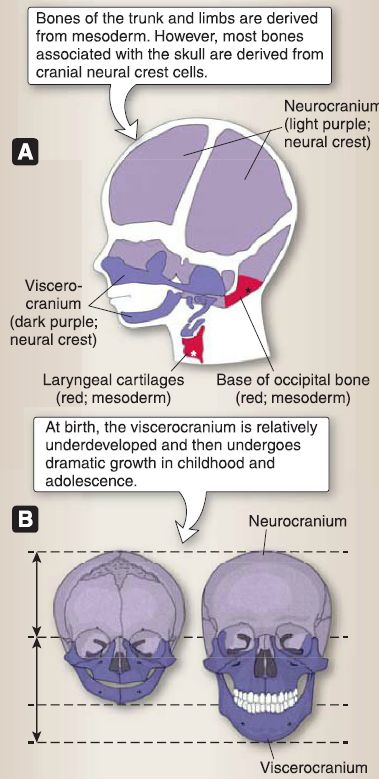
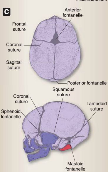
Figure 2: A, Newborn skull. B, Postnatal growth of skull. C, Sutures and fontanelles.
a. Sutures: During fetal life and infancy, the flat bones of the skull are separated by dense connective tissue (fibrous joints) called sutures. There are five sutures: frontal, sagittal, lambdoid, coronal, and squamous sutures. Sutures allow the flat bones of the skull to deform during childbirth (called molding) and to expand during childhood as the brain grows. Typically, the frontal suture closes by the eighth year of life, while the others slowly close into adulthood.
b. Fontanelles: Fontanelles are large, fibrous areas where several sutures meet. There are six fontanelles: anterior, posterior, two sphenoid, and two mastoid fontanelles. The anterior fontanelle is the largest fontanel and is readily palpable in the infant. The anterior fontanel pulsates because of the underlying cerebral arteries and can be used to obtain a blood sample from the underlying superior sagittal sinus. The anterior fontanelle and the mastoid fontanelles close at about age 2 years when the main growth of the brain ceases. The posterior fontanelle and the sphenoid fontanelles close at about age 6 months.
2. Adult skull: The adult bony skull can be divided into two parts: the neurocranium and the viscerocranium. Figures 3 through 5 provide various views of the skull.
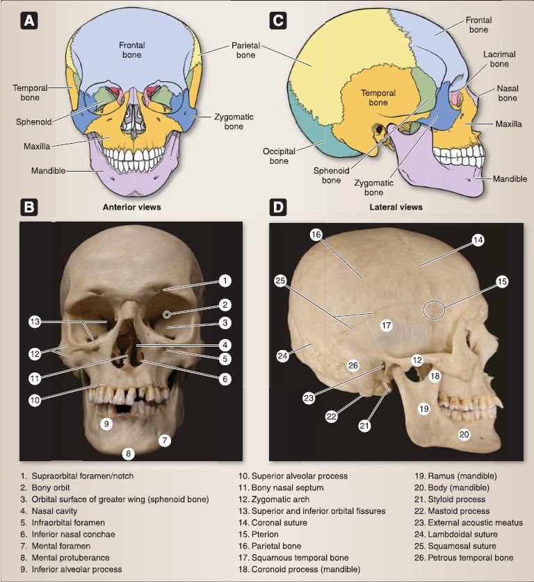
Figure 3 : Adult cranium. A and C, Color-coded bones. B and D, Skeletal specimens.
a. Neurocranium: Collectively, the neurocranium is made up of frontal, paired parietal, paired temporal, ethmoid, sphenoid, and occipital bones. The superior portions of the frontal, parietal, and occipital bones contribute to the formation of the calvaria (skull cap).
[1] Calvaria: Distinguishing features of the calvaria include the sagittal, coronal, and lambdoid sutures, previously described. The junctions of the sagittal suture with the coronal and lambdoidal sutures are termed bregma and lambda, respectively. These junctions represent the closure sites of the anterior and posterior fontanelles, respectively.
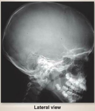
Figure 4: Plain film radiograph (child skull, face and upper cervical spine).
(a) Vertex: The superiormost point of the calvaria is the vertex, located approximately midway along the sagittal suture.
(b) Temporal fossa: Laterally, the pterion represents the junction of the frontal, parietal, sphenoid, and temporal (squamosal) bones and is situated in a region called the temporal fossa. Superior and inferior temporal lines serve as superior and posterior boundaries of this region, which is comprised of mainly by the parietal and temporal (squamous) bones, with anterior contributions from the sphenoid (greater wing) and frontal bones.
[2] Base: The frontal, ethmoid, sphenoid, petrous temporal, and occipital bones form the cranial base.
(a) Fossae: Internally, the cranial base is further divided into anterior, middle, and posterior fossae :
(i) Anterior fossa: Frontal, ethmoid, and sphenoid bones contribute to the anterior fossa.
(ii) Middle fossa: Sphenoid and temporal bones contribute to the middle fossa.
(iii) Posterior fossa: Sphenoid and occipital bones contribute to the posterior fossa.
(b) Foramina and fissures: Bilateral foramina and fissures are present in each fossa, allowing for the passage of important neurovascular structures (Table 1; also see Fig. 5). Additional foramina occur throughout the base of the skull to transmit emissary veins, including the foramen cecum (frontal) and condylar and mastoid foramina (occipital).
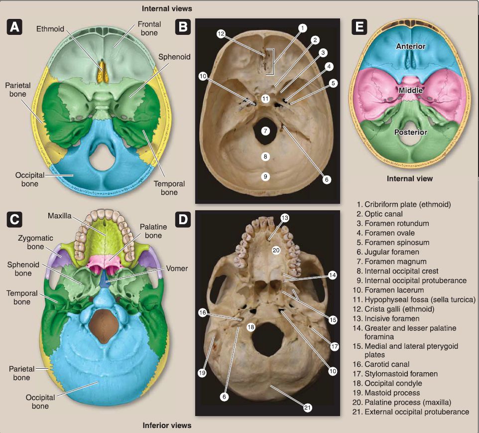 Figure 5: Adult cranium. A and C, Color-coded bones. Band D, Skeletal specimens. E, Cranial fossae.
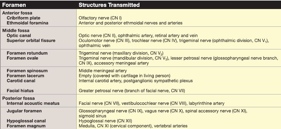
Table 1: Internal Neurocranial Foramina and Contents
(c) External surface features: Inferiorly, the external cranial base has several surface features that serve as attachment sites for muscles and fascia as well as regions of bony articulations. Of particular importance are the paired occipital condyles, superior sites for articulation with C1 (atlas) to form the atlanto-occipital joint. The mastoid and styloid processes (petrous temporal) are located laterally to the occipital condyles. Many of the previously mentioned foramina are visible inferiorly, and additional foramina include the opening of the incisive canal (maxilla), greater and lesser palatine foramina (palatine), stylomastoid foramen (petrous temporal), and the entry site for the carotid canal (petrous temporal) (see Table 2; also see Fig.).
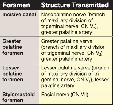
Table 2: Inferior Neurocranial Foramina and Contents
(d) Occipital bone: Externally, the occipital bone represents the majority of the posterior portion of the skull, with additional contribution from the parietal bones superiorly and the mastoid processes (petrous temporal) inferiolaterally.
Posteriorly, the occipital bone is characterized by a midline palpable elevation of bone called the external occipital protuberance. The superior nuchal line extends laterally from the protuberance, serving as the superiormost attachment site for neck structures.
The less prominent inferior nuchal line lies inferior to the protuberance.
b. Viscerocranium: Located anteriorly, the viscerocranium represents the bony scaffold of the face and is made up of bones that surround and form three main facial regions-orbit, nose, and mouth. Bones that contribute to the formation of the viscerocranium include parasagittal paired maxillae, zygomatic, nasal, lacrimal, and inferior turbinate (nasal concha) bones and midline singular mandible, ethmoid, and vomer bones (see Figs. 3 and 5).
[1] Frontal bone: This bone forms the majority of the forehead of the face and contributes to the superior rim (supraorbital margin) of the orbit. Bilateral supraorbital notches are found medially along the supraorbital margins. A smooth, midline depression, the glabella, sits between the paired supraciliary arches and is easily palpated between the eyebrows, superior to the nasal bridge-formed by paired nasal bones.
[2] Zygomatic and maxillary bones: These bones complete the orbital margin inferiolaterally and inferomedially, respectively. Each zygomatic bone has a zygomaticotemporal foramen anterior to the temporal process of the zygomatic bone. This process articulates with the zygomatic process of the temporal bone to form the zygomatic arch. The external acoustic meatus (petrous temporal) lies just posterior to the zygomatic arch. Bilateral infraorbital foramina are located just inferior to the infraorbital margin on the superior aspect of the maxilla. The intermaxillary suture represents the fusion site for the paired maxillae, forming the upper jaw and characterized by alveolar processes that support the upper (maxillary) teeth.
[3] Mandible: The jaw is completed inferiorly by the Ushaped mandible, also characterized by alveolar processes that support the lower (mandibular) teeth. A midline mental protuberance is found inferiorly and is framed by bilateral mental foramina. Laterally, the mandible is characterized by a body, angle, and ramus.
Superior to the ramus, the coronoid process and head of the mandible are situated anteriorly and posteriorly, respectively. The head of the mandible articulates with the mandibular fossa (petrous temporal) to form the temporomandibular joint.
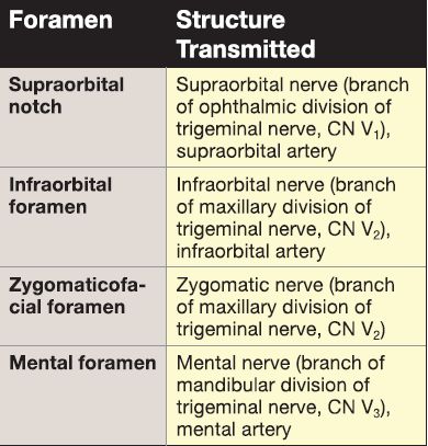
[4] Foramina: Refer to Table 3 for additional details about viscerocranial foramina.
The scalp is a multilayer structure that covers the neurocranium, spanning between the supraorbital margins anteriorly to the superior nuchal lines posteriorly and extending laterally to the zygomatic arches.
Table 3: Viscerocranial Foramina and Contents
1. Scalp proper: The first three layers of the scalp are called the scalp proper and comprise skin, connective tissue, and aponeurosis. The skin of the scalp has a rich blood supply and contains multiple sweat and sebaceous glands and hair follicles. The thick subcutaneous connective tissue layer also has a substantial neuromuscular supply.
The aponeurosis layer provides a tendon-like bridge between the anterior and posterior bellies of the occipitofrontalis muscle and also serves as an attachment site for the superior auricular muscle (Fig. 6A).
2. Loose connective tissue layer: Deep to the aponeurosis, the loose connective tissue layer allows for movement of the scalp proper on the skull and contains emissary veins.
3. Pericranium: Finally, the innermost layer, the pericranium, is a dense connective tissue layer that is firmly attached to the calvaria, forming the periosteum.
4. Innervation: The skin of the scalp is innervated by branches of the trigeminal nerve and cervical anterior and posterior rami (see Fig.6B).
a. Anterior and superior scalp: This area is innervated by the supratrochlear and infratrochlear nerves (CN V1, ophthalmic).
b. Anterolateral scalp: This area is innervated by the zygomaticotemporal (CN V2, maxillary) and auriculotemporal (CN V3 ,mandibular) nerves.
c. Posterolateral scalp: This area is innervated by the lesser occipital nerve (anterior rami C2 , C3).
d. Posterior and superior scalp: This area is innervated by the greater occipital nerve (posterior rami C2).
5. Vasculature: The scalp receives blood supply primarily from branches of the external carotid artery, including posterior auricular, occipital, and superficial temporal arteries. The anterior scalp is supplied by vessels that indirectly arise from the internal carotid artery, including the supratrochlear and supraorbital arteries.
Typically, veins of the same name drain the superficial scalp. There are no lymph nodes in the superficial scalp, so all lymph is drained into the superficial ring of lymph nodes located at the intersection of the head and neck.
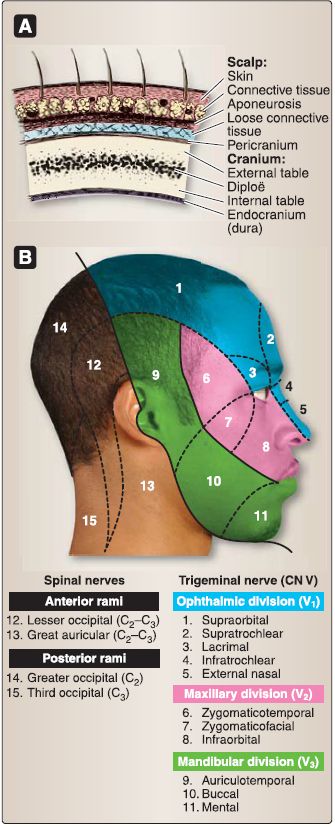
Figure 6: Scalp. A, Layers. B, Innervation of the scalp and face.


