
Peritoneal Cavity
 المؤلف:
Kelly M. Harrell and Ronald Dudek
المؤلف:
Kelly M. Harrell and Ronald Dudek
 المصدر:
Lippincott Illustrated Reviews: Anatomy
المصدر:
Lippincott Illustrated Reviews: Anatomy
 الجزء والصفحة:
 15-7-2021
 11670
11670
Peritoneal Cavity
The peritoneal cavity is a potential space between parietal and visceral peritoneal layers that line the inner surface of the abdominal body wall and abdominal organs (viscera), respectively (Fig. 1).
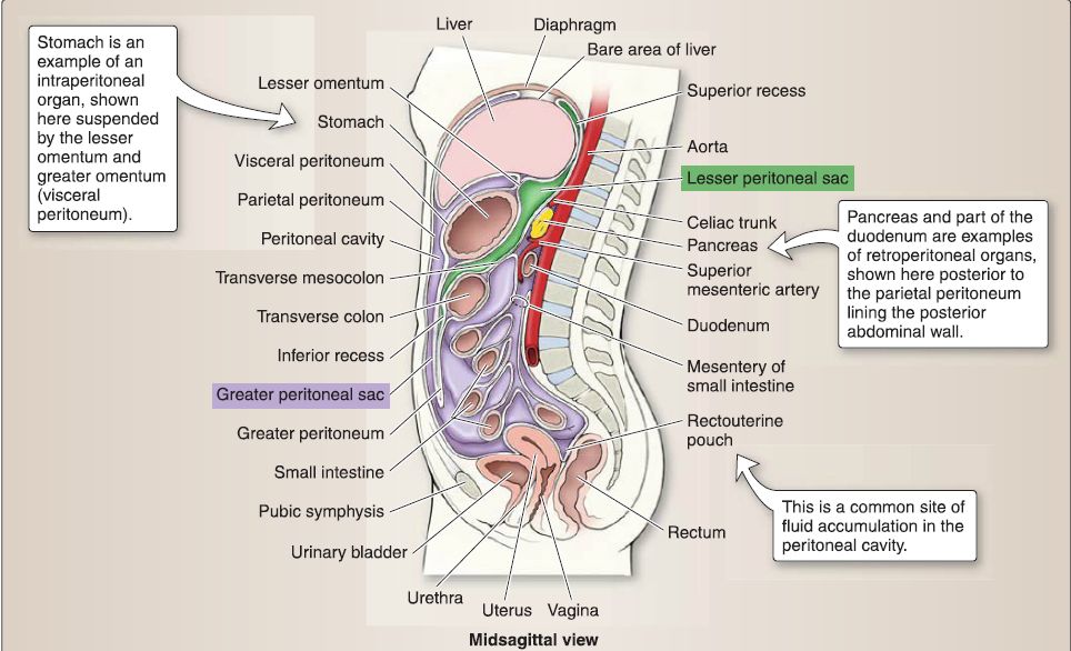 Figure 1: Peritoneal cavity.
A. Development
Recall that during the fourth week of development, the intraembryonic coelom has developed and is being partitioned into pleural, pericardia, and peritoneal cavities . Pleuroperitoneal folds expand medially to begin to close off the right and left pericardioperitoneal canals. During this process, the folds thin to become pleuroperitoneal membranes, which will eventually fuse with the septum transversum and dorsal mesentery of the esophagus. This fusion, along with contributions from lateral body wall ingrowth, forms the primitive thoracic diaphragm and partitions the thoracic and abdominal cavities.
1. Mesentery: Lateral mesoderm contributes to the development of serous membranes that make up the layers of the peritoneal cavityparietal and visceral peritoneum. At the junction of parietal and visceral peritoneum, a double layer of peritoneum, called mesentery, exists to suspend or cover viscera and provide a passageway for neurovascular structures to reach abdominal viscera. In the developing human, primitive ventral and dorsal mesenteries (mesogastria) connect the gut tube to the ventral or dorsal body wall, respectively.
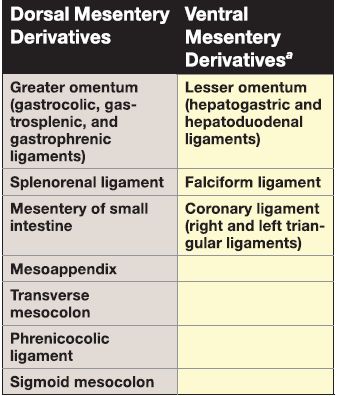
In the early stages of development, this arrangement partitions the abdominal cavity into right and left halves. As gut tube development and rotation proceeds, the ventral mesentery remains only in the area of the liver, proximal duodenum, and lesser curvature of the stomach. The dorsal mesentery develops into visceral peritoneum associated with the greater curvature of the stomach, portions of the small and large intestines, spleen, kidney, and diaphragm (Table 1).
Table 1: Dorsal and Ventral Mesentery Derivatives
Final positioning of the ventral and dorsal mesenteries contributes to the formation of a small space posterior to the stomach called the lesser peritoneal sac (omental bursa), which has limited communication with a larger space that occupies most of the peritoneal cavity called the greater peritoneal sac.
2. Parietal peritoneum: With the elongation of the trunk, the final position of abdominal contents extends into the true pelvis. Parietal peritoneum also extends into the true pelvis, thus serving as a drape over pelvic viscera. This draping of parietal peritoneum completely closes off the peritoneal cavity in males but allows for communication externally in females by way of the uterine tubes, which are not fully covered by parietal peritoneum .
B. Anatomy
The peritoneal cavity is formed during development by primitive dorsal and ventral mesenteries. Following birth, the primitive mesenteries are designated as either an omentum or a "ligament" and are named by the structures they span between or surround. Viscera that are suspended in these peritoneal structures are described as intraperitoneal, whereas viscera that are only partially covered (one side) by peritoneum and posterior to the parietal peritoneum are described as retroperitoneal. These relationships between peritoneum and viscera occur as a result of GI tract development (Table 2 and Fig. 2).
Table 2: lntraperitoneal and Retroperitoneal Viscera
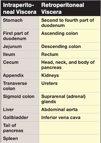
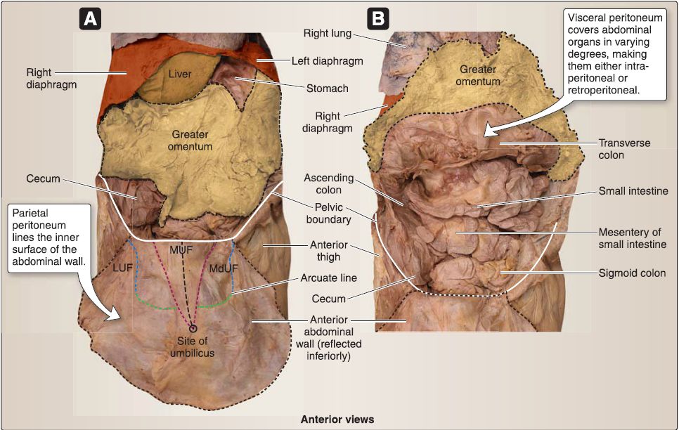
Figure 2: Peritoneal structures. Open abdominal cavity. A, Greater omentum in position. B, Greater omentum reflected superiorly. LUF = lateral umbilical fold, MdUF = medial umbilical fold, MUF = median umbilical fold.
1. Greater omentum: The greater omentum is apron-like in appearance and contains variable amounts of fat. Arising from the greater curvature of the stomach, it drapes anterior to the transverse colon and-in varying degrees-the small intestines before doubling back and ascending to attach to the transverse colon. It is further divided into different portions, including the gastrocolic ligament (attached to transverse colon), gastrosplenic ligament (attached to spleen hilum), and gastrophrenic ligament (attached to abdominal surface of the diaphragm).
2. Lesser omentum: The lesser omentum spans between the lesser curvature of the stomach and the first part of the duodenum to the liver. Although the lesser omentum is continuous, it is further distinguished by these attachments into two ligaments-hepatogastric and hepatoduodenal. The lesser omentum closes off the lesser peritoneal sac anteriorly, and the free edge of the hepatoduodenal ligament serves as a boundary of the omental foramen (foramen of Winslow). The hepatoduodenal ligament also contains the portal triad (common bile duct, proper hepatic artery, and hepatic portal vein), as shown in Figure 3.
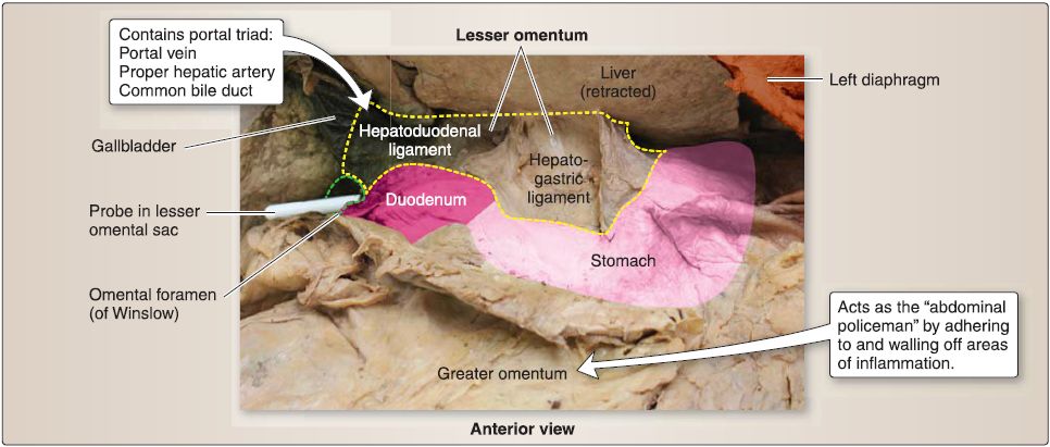
Figure 3: Lesser omentum and sac.
3. Splenorenal ligament: This ligament spans between the hilum of the spleen and the left kidney. The splenic artery travels through this ligament and gives off important branches to portions of the stomach.
4. Small intestine mesentery: This fat-laden, fan-shaped mesentery extends from the posterior abdominal wall to suspend the jejunum and ileum. Intestinal vessels and autonomic nerves travel through the mesentery substance to reach the organ walls.
5. Transverse mesocolon: This ligament suspends the transverse colon from the posterior abdominal wall and spans across the descending (second part) duodenum and the majority of the pancreas. The gastrocolic ligament is positioned superior and anterior to the transverse mesocolon.
6. Phrenicocolic ligament: This ligament spans between the left colic (splenic) flexure of the large intestine and the abdominal surface of the left hemidiaphragm. These connections form a "shelf" for the spleen.
7. Sigmoid mesocolon: This ligament spans between the inferior, left posterior abdominal wall and the sigmoid colon.
8. Mesoappendix: Small in nature, this mesentery spans between the posterior abdominal wall and appendix.
9. Falciform ligament: This thin fold of mesentery extends from the anterior liver surface to the internal surface of the anterior abdominal wall. It is sickle-shaped, and its inferior border houses the ligamentum teres hepatis (round ligament of the liver), which represents the obliterated umbilical vein.
10. Coronary ligaments: A divergence occurs at the superior limit of the falciform ligament on the surface of the liver. This separation of visceral peritoneal layers forms the right and left coronary ligaments. At their most distal extensions, the right and left coronary ligaments reverse in direction to form the right and left triangular ligaments, respectively. Collectively, these reflections surround an area on the liver-the bare area-that lacks visceral peritoneum.
C. Peritoneal sacs
The peritoneal cavity is described as having two sacs-lesser and greater. The lesser and greater peritoneal sacs communicate by way of the omental foramen (foreman of Winslow). The omental foramen is bound posteriorly by the inferior vena cava (IVG) and anteriorly by the portal triad (see Fig. 3 ).
1. Lesser sac: The lesser peritoneal sac (omental bursa) allows for movement of the stomach on posterior structures. The boundaries of the lesser peritoneal sac are as follows.
a. Anterior: The liver, stomach, and lesser omentum (hepatogastric and hepatoduodenal ligaments) form the anterior border.
b. Posterior: The diaphragm forms the posterior border.
c. Right: The right border is formed by the liver.
d. Left: Gastrosplenic and splenorenal ligaments form the left border.
2. Greater sac: The greater peritoneal sac occupies the remaining space in the abdomen spanning from the diaphragm to the pelvis. A series of spaces (pouches, recesses, paracolic "gutters") occur within the greater peritoneal sac, allowing for normal circulation as well as accumulation of peritoneal fluids in pathologic states .
 الاكثر قراءة في علم التشريح
 اخر الاخبار
اخبار العتبة العباسية المقدسة


