

النبات

مواضيع عامة في علم النبات

الجذور - السيقان - الأوراق

النباتات الوعائية واللاوعائية

البذور (مغطاة البذور - عاريات البذور)

الطحالب

النباتات الطبية


الحيوان

مواضيع عامة في علم الحيوان

علم التشريح

التنوع الإحيائي

البايلوجيا الخلوية


الأحياء المجهرية

البكتيريا

الفطريات

الطفيليات

الفايروسات


علم الأمراض

الاورام

الامراض الوراثية

الامراض المناعية

الامراض المدارية

اضطرابات الدورة الدموية

مواضيع عامة في علم الامراض

الحشرات


التقانة الإحيائية

مواضيع عامة في التقانة الإحيائية


التقنية الحيوية المكروبية

التقنية الحيوية والميكروبات

الفعاليات الحيوية

وراثة الاحياء المجهرية

تصنيف الاحياء المجهرية

الاحياء المجهرية في الطبيعة

أيض الاجهاد

التقنية الحيوية والبيئة

التقنية الحيوية والطب

التقنية الحيوية والزراعة

التقنية الحيوية والصناعة

التقنية الحيوية والطاقة

البحار والطحالب الصغيرة

عزل البروتين

هندسة الجينات


التقنية الحياتية النانوية

مفاهيم التقنية الحيوية النانوية

التراكيب النانوية والمجاهر المستخدمة في رؤيتها

تصنيع وتخليق المواد النانوية

تطبيقات التقنية النانوية والحيوية النانوية

الرقائق والمتحسسات الحيوية

المصفوفات المجهرية وحاسوب الدنا

اللقاحات

البيئة والتلوث


علم الأجنة

اعضاء التكاثر وتشكل الاعراس

الاخصاب

التشطر

العصيبة وتشكل الجسيدات

تشكل اللواحق الجنينية

تكون المعيدة وظهور الطبقات الجنينية

مقدمة لعلم الاجنة


الأحياء الجزيئي

مواضيع عامة في الاحياء الجزيئي


علم وظائف الأعضاء


الغدد

مواضيع عامة في الغدد

الغدد الصم و هرموناتها

الجسم تحت السريري

الغدة النخامية

الغدة الكظرية

الغدة التناسلية

الغدة الدرقية والجار الدرقية

الغدة البنكرياسية

الغدة الصنوبرية

مواضيع عامة في علم وظائف الاعضاء

الخلية الحيوانية

الجهاز العصبي

أعضاء الحس

الجهاز العضلي

السوائل الجسمية

الجهاز الدوري والليمف

الجهاز التنفسي

الجهاز الهضمي

الجهاز البولي


المضادات الميكروبية

مواضيع عامة في المضادات الميكروبية

مضادات البكتيريا

مضادات الفطريات

مضادات الطفيليات

مضادات الفايروسات

علم الخلية

الوراثة

الأحياء العامة

المناعة

التحليلات المرضية

الكيمياء الحيوية

مواضيع متنوعة أخرى

الانزيمات
Osteology
المؤلف:
Kelly M. Harrell and Ronald Dudek
المصدر:
Lippincott Illustrated Reviews: Anatomy
الجزء والصفحة:
10-7-2021
4110
Osteology
Back stability is provided by the vertebral column or spine , which isdivided into cervical, thoracic, lumbar, sacral, and occygeal regions. As shown in Figure 1, the adult vertebral column typically contains 33 vertebrae: 7 cervical, 12 thoracic, 5 lumbar:5 , 5 sacral (fused), and 3-5 coccygeal (fused). Vertebrae are separated by fibrocartilaginous intervertebral discs and united through a series of joint , igaments, and capsules. Functionally, the vertebral column serves to protect the spinal cord and spinal nerves and support body weight superior to the pelvis. It also allows for varying degrees of motion arnsi plays an integral part in posture and gait. Most spinal motion occurs superior to the sacral level and is further dictated by articular orientation and bony attachments (e.g., ribs in the thoracic region). Spinal motion includes extension, flexion, lateral side-bending, rotation, and circumduction.
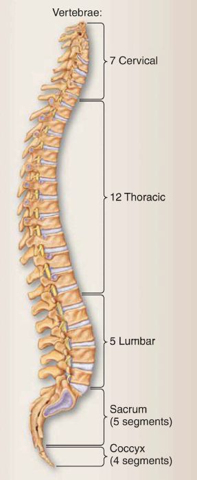
Figure 1:Vertebral column.
A. Vertebrae
A vertebra is generally arranged as a weight-bearing body anteriorly, a vertebral (neural) arch and spinous process posteriorly, and transverse processes laterally. Articular processes project superiorly and inferiorly (Fig. 2).
1. Vertebral body: The vertebral body is the weight-bearing portion of the vertebra and also serves as the site for intervertebral disc attachment. Body size and shape vary throughout the length of the vertebral column.
2. Vertebral arch: The vertebral arch protects the spinal cord and is formed by two pedicles that project posteriorly from the body and two laminae that meet in the mid line at the spinous process.
3. Vertebral foramen: The vertebral canal houses the spinal cord and is formed by the collection of vertebral foramen along the length of the spine. Right and left sets of superior and inferior articular processes project from the junction of the pedicles and laminae.

Figure 2: General vertebral structures.
4. Vertebral notches: Laterally, superior and inferior vertebral notches present as indentations posterior to the vertebral body.
a. lntervertebral foramina: The inferior and superior vertebral notches of adjacent vertebrae combine to form paired intervertebral foramina at each spinal level. Spinal nerves and vasculature travel through intervertebral foramina on each side of the column.
b. Sacral foramina: In the sacral region, spinal nerves exit the canal through anterior and posterior sacral foramina.
5. Processes: Muscles and ligaments attach to both spinous and transverse processes. Transverse processes in the thoracic region also articulate with ribs posteriorly and support the thoracic cage.
B. Regional characteristics
Regionally, vertebrae have distinct structural characteristics that further dictate function. For example, articular processes are oriented in different planes to allow or limit spinal mobility. Variations in size and shape can also allow for transmission of neural structures, such as the cervical and lumbar spinal cord enlargements, in respective regions.
1. Cervical (C1-C7): As shown in Figures 3 and 4, cervical vertebrae feature a small body and a large, triangular vertebral foramen. A foramen transversarium in the transverse processes accommodates vertebral vessels and sympathetic plexuses. Articular processes are oriented in the horizontal (transverse) plane. Spinous processes in C3-C5 are short and bifid (divided into two equal parts). Unique cervical vertebrae are described as follows.
a. C1 (atlas): This ring-like vertebra has neither body nor spinous process. It articulates with occipital condyles through two lateral masses (atlanto-occipital joint) and has a small anterior facet for articulation with the dens (odontoid process) of C2.
b. C2 (axis): This vertebra is strong, featuring a dens that projects superiorly to articulate with C1 (pivot joint).
c. C7 (vertebra prominens): This vertebra with its long, prominent spinous process makes a reliable surface landmark.

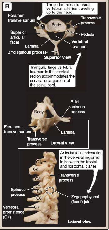
Figure 3: A, Atlas (C1) and axis (C2). B, Cervical vertebrae features.
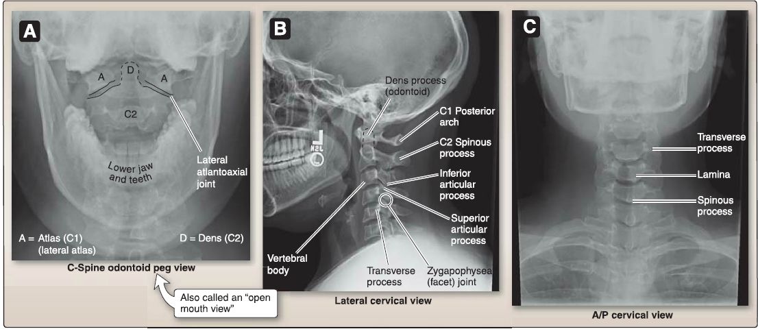
Figure 4: Cervical plain film views (A-C).
2. Thoracic (T1-T12}: As shown in Figures 5 and 6, thoracic vertebrae feature a heart-shaped body with costal demifacets for articulation with the heads of ribs and a small, circular vertebral foramen. In T1-T10 vertebrae, transverse processes have costal facets for articulation with rib tubercles, articular processes oriented in the frontal (coronal) plane, and long posteroinferiorly sloping, overlapping spinous process.
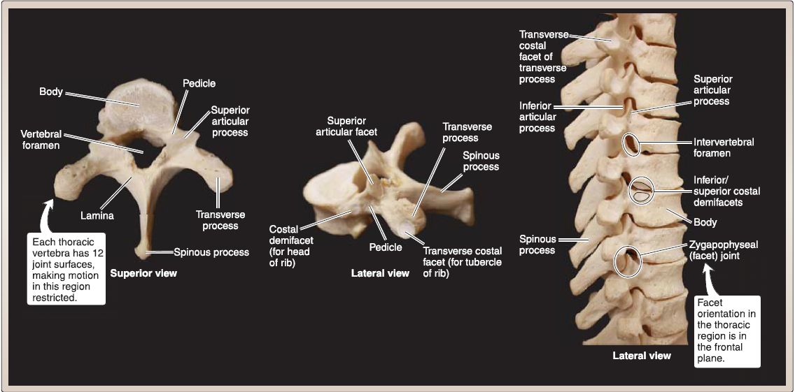
Figure 5 :Thoracic regional features.
 Figure 6: Thoracic plain film views (A and B).
3. Lumbar (L 1-L5): As shown in Figures 7 and 8, lumbar vertebrae have large, kidney-shaped bodies and large, triangular vertebral foramina. Their articular facets are oriented in the sagittal plane, and they have short, hatched-shaped spinous processes. A mammillary process is located on the superior articular process.
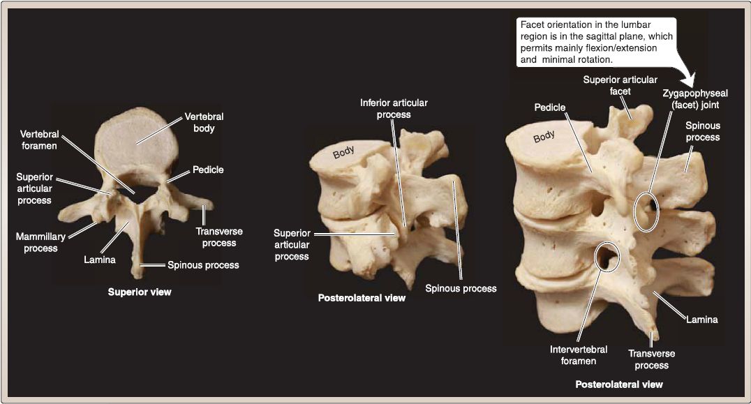
Figure 7:Lumbar regional features.
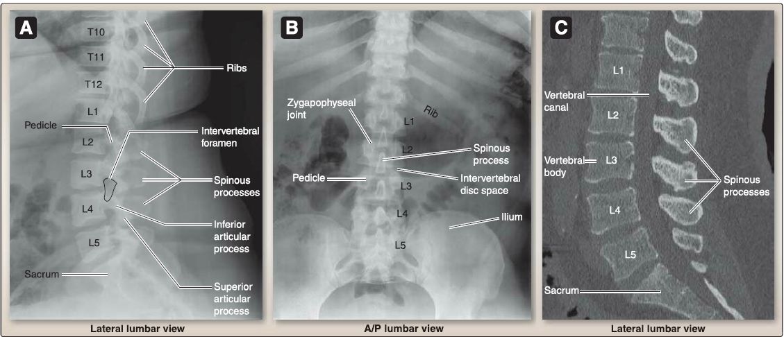
Figure 8 :Plain film, A/8 and computed tomography (CT), C.
4. Sacral (S1-S5): This fused, wedge-shaped bone is concave anteriorly and convex posteriorly. It features anterior and posterior foramina and a sacral canal and hiatus (Fig. 9).
5. Coccygeal: This beak-like bone comprises three to five small, fused vertebrae. It is a tail-like structure at the caudalmost region of the vertebral column.
 Figure 9:Sacral and coccygeal regional features.
C. lntervertebral discs
As shown in Figure 10, fibrocartilage intervertebral discs are arranged between all presacral vertebrae, except for between the occiput, atlas (C1), and axis (C2). In the adult, the discs constitute -25% of the total length of the spine.
1. Composition: Each disc consists of an outer annulus fibrosis and an inner nucleus pulposus. The strong outer annulus is made up of 6-10 fibrocartilaginous rings {lamellae) arranged concentrically at opposite angles. The inner nucleus pulposus is a gel-like connective tissue structure with high water content. Collectively, these structures permit spinal movement and transmit loads across each vertebral segment, in addition to binding vertebral bodies together.
2. Regional characteristics: Regionally, intervertebral discs vary in shape and thickness. Variations in thickness permit different ranges of motion, while shape is dictated mainly by secondary spinal curvatures (see 11.D). For example, in the cervical and lumbar regions, discs are thicker anteriorly, whereas thickness is generally uniform throughout the thoracic spine.

Figure 10 : lntervertebral disc.
D. Curvatures
In the adult, there are four distinct spinal curvatures, which are described as being either primary or secondary (Fig. 2.11 ). Primary curvatures include those in the thoracic and sacral regions. These are concave anteriorly and represent curvatures that develop during the fetal period. Secondary curvatures are found in the cervical and lumbar regions and are concave posteriorly. Cervical and lumbar curvatures develop as an infant begins to hold his or her head erect and walk, respectively. Deviations from normal spinal curvatures may lead to clinical impairments in the back.
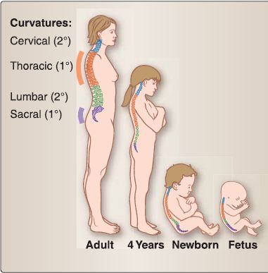
Figure 11: Primary (1 °) and secondary (2°) spine curvatures. Normal curvatures of the
spine from fetus to adult.
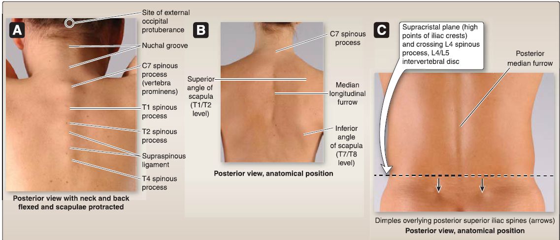
Figure 12: Back surface anatomy (A-C).
E. Surface anatomy
Clinicians need to have a solid understanding of back surface anatomy to detect abnormalities (Fig. 12A-C). Along the midline of the back is a recess called the posterior median furrow, which overlies vertebral spinous processes. Spinous processes project posteriorly and are best observed when a patient's spine is in a flexed position.
1. Cervical region: The spinous process of C7 (vertebra prominens) is often the most visible in this region. The spinous process of T1 can also be very distinct and easily mistaken for C7. Superior to Cl, a thick nuchal ligament overlies the cervical spinous processes of C2-C6, making it difficult but not impossible to palpate at these levels.
2. Thoracic region: Here, spinous processes are longer and slope inferiorly. Because of this arrangement, palpation of a thoracic spinous process does not always correspond to vertebral body level. The superior angle of the scapula typically corresponds to the T1/T2 spinous process level, while the inferior angle of the scapula lies at the T7 /T8 level.
3. Lumbar region: Lumbar spinous processes are large and easily palpated with the patient in a flexed position. The L4 spinousprocess lies at the level of the iliac crest, which is an important landmark for spinal anesthesia and lumbar puncture procedures.
4. Sacral region: Sacral spines of the first three fused segments can be palpated. Commonly, a small dimple representing the posterior superior iliac spine can be visualized at the level of the S2 spinous process.
 الاكثر قراءة في علم التشريح
 اخر الاخبار
اخبار العتبة العباسية المقدسة

الآخبار الصحية


 قسم الشؤون الفكرية يصدر كتاباً يوثق تاريخ السدانة في العتبة العباسية المقدسة
 "المهمة".. إصدار قصصي يوثّق القصص الفائزة في مسابقة فتوى الدفاع المقدسة للقصة القصيرة
 (نوافذ).. إصدار أدبي يوثق القصص الفائزة في مسابقة الإمام العسكري (عليه السلام)


