


النبات

مواضيع عامة في علم النبات
الجذور - السيقان - الأوراق
النباتات الوعائية واللاوعائية
البذور (مغطاة البذور - عاريات البذور)
الطحالب
النباتات الطبية
الحيوان
مواضيع عامة في علم الحيوان
علم التشريح
التنوع الإحيائي
البايلوجيا الخلوية
الأحياء المجهرية
البكتيريا
الفطريات
الطفيليات
الفايروسات
علم الأمراض
الاورام
الامراض الوراثية
الامراض المناعية
الامراض المدارية
اضطرابات الدورة الدموية
مواضيع عامة في علم الامراض
الحشرات
التقانة الإحيائية
مواضيع عامة في التقانة الإحيائية
التقنية الحيوية المكروبية
التقنية الحيوية والميكروبات
الفعاليات الحيوية
وراثة الاحياء المجهرية
تصنيف الاحياء المجهرية
الاحياء المجهرية في الطبيعة
أيض الاجهاد
التقنية الحيوية والبيئة
التقنية الحيوية والطب
التقنية الحيوية والزراعة
التقنية الحيوية والصناعة
التقنية الحيوية والطاقة
البحار والطحالب الصغيرة
عزل البروتين
هندسة الجينات
التقنية الحياتية النانوية
مفاهيم التقنية الحيوية النانوية
التراكيب النانوية والمجاهر المستخدمة في رؤيتها
تصنيع وتخليق المواد النانوية
تطبيقات التقنية النانوية والحيوية النانوية
الرقائق والمتحسسات الحيوية
المصفوفات المجهرية وحاسوب الدنا
اللقاحات
البيئة والتلوث
علم الأجنة
اعضاء التكاثر وتشكل الاعراس
الاخصاب
التشطر
العصيبة وتشكل الجسيدات
تشكل اللواحق الجنينية
تكون المعيدة وظهور الطبقات الجنينية
مقدمة لعلم الاجنة
الأحياء الجزيئي
مواضيع عامة في الاحياء الجزيئي
علم وظائف الأعضاء
الغدد
مواضيع عامة في الغدد
الغدد الصم و هرموناتها
الجسم تحت السريري
الغدة النخامية
الغدة الكظرية
الغدة التناسلية
الغدة الدرقية والجار الدرقية
الغدة البنكرياسية
الغدة الصنوبرية
مواضيع عامة في علم وظائف الاعضاء
الخلية الحيوانية
الجهاز العصبي
أعضاء الحس
الجهاز العضلي
السوائل الجسمية
الجهاز الدوري والليمف
الجهاز التنفسي
الجهاز الهضمي
الجهاز البولي
المضادات الميكروبية
مواضيع عامة في المضادات الميكروبية
مضادات البكتيريا
مضادات الفطريات
مضادات الطفيليات
مضادات الفايروسات
علم الخلية
الوراثة
الأحياء العامة
المناعة
التحليلات المرضية
الكيمياء الحيوية
مواضيع متنوعة أخرى
الانزيمات

Nervous System
 المؤلف:
Kelly M. Harrell and Ronald Dudek
المؤلف:
Kelly M. Harrell and Ronald Dudek
 المصدر:
Lippincott Illustrated Reviews: Anatomy
المصدر:
Lippincott Illustrated Reviews: Anatomy
 الجزء والصفحة:
 10-7-2021
 4276
4276


+
-
20
Nervous System
The nervous system can be anatomically divided into the central nervous system (CNS) and the peripheral nervous system (PNS). The nervous system can also be functionally divided into the somatic nervous system, which controls voluntary activities by innervating skeletal muscle, and the autonomic (or visceral) nervous system (ANS), which controls involuntary activities by innervating viscera, smooth muscle, cardiac muscle, and glands.
A. Neuron
The neuron is the structural and functional unit of the nervous system. It has the capacity to receive impulses from other neurons or from receptor organs (i.e., a sensory function) as well as to transmit impulses to other neurons or to effector organs (i.e., a motor function). A neuron consists of a perikaryon (soma), dendrites, and an axon (Figs. 1 and 2). The perikaryon is the cell body of the neuron and contains the nucleus along with the organelles. The dendrites receive information from other neurons or from the environment and transmit the information toward the perikaryon. The axon generates, propagates, and transmits an action potential.
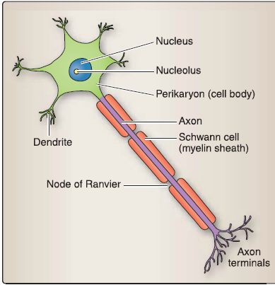
Figure 1: Neuron.
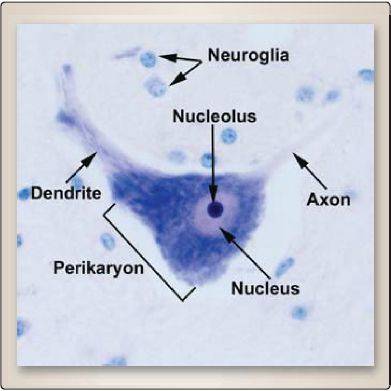
Figure 2 :Light micrograph of a neuron.
B. Synapse
A synapse is a specialized junction by which neurons communicate with one another. The three main types are the axodendritic synapse between an axon and a dendrite, the axosomatic synapse between an axon and the perikaryon (soma), and the axoaxonic synapse between an axon and another axon (Fig. 3). Synapses consist of pre- and postsynaptic components and the synaptic cleft (Fig. 4).
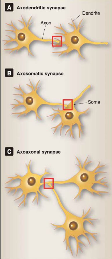
Figure 3 : A to C, Types of synapses.
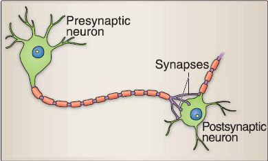
Figure 4 : Pre- and postsynaptic neurons.
1. Presynaptic: This component is characterized morphologically by the presence of 40- to 60-nm-diameter synaptic vesicles that contain a neurotransmitter. The presynaptic cell membrane contains voltage-gated Ca2+ channels that open when a depolarization reaches the synaptic terminal and that allow an influx of Ca2+ from the extracellular milieu. The influx of Ca2+ causes the docking of synaptic vesicles at the presynaptic membrane and the release of neurotransmitter into the synaptic cleft (Fig. 5). The synaptic cleft is a 20- to 30-nm space between the presynaptic and postsynaptic membranes. This is where the neurotransmitter is released into in order to bind to its receptor on the postsynaptic membrane.
2. Postsynaptic: This component is characterized morphologically by the presence of the postsynaptic density located just beneath the postsynaptic cell membrane (see Fig. 5). The postsynaptic cell membrane contains either transmitter-gated ion channels or G protein-linked receptors that bind a specific neurotransmitter.

Figure 5 : Synapse.
C. Central Nervous System
The Central nervous system (CNS) consists of the brain and the spinal cord. When examining the brain or spinal cord with the unaided eye, the white matter and gray matter can be distinguished (Fig. 6).
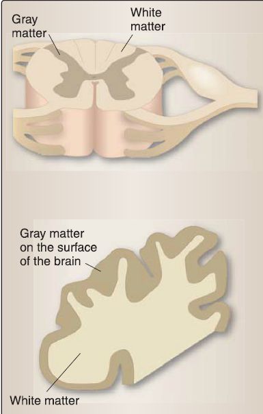
Figure 6 :White and gray matter.
The appearance of the white matter is due to the abundance of myelinated axons, whereas the appearance of the gray matter is due to the scarcity of myelinated axons. The neuropil is a broad term used to describe the unmyelinated and myelinated axons that form a synapse-rich meshwork in the gray matter, "and includes dendrites and neuroglial cell processes".
1. Cells: CNS neuroglia include astrocytes, oligodendrocytes,microglia, and ependymal cells (Fig. 7). The astrocyte has numerous functions, some of which include 1) projects foot processes to capillaries that contribute to the blood-brain barrier, 2) plays a role in the metabolism of neurotransmitters, and 3) buffers the [K+] of the CNS extracellular space. The oligodendrocyte produces a myelin sheath that surrounds and insulates a portion of an axon. The junction between adjacent oligodendrocyte processes on an axon is called the node of Ranvier. The microglia have a phagocytic function and proliferate in regions of injury or disease whereby they are then called reactive microglia. The ependymal cells line the ventricles of the brain and the central canal of the spinal cord.

Figure 7: Neuroglia. A, Oligodendrocytes and Schwann cells. B, Astrocytes.C, Ependyma and microglia.
2. Meninges: CNS connective tissue components called the meninges protect the underlying brain and spinal cord; provide a framework for arteries, veins, and dural sinuses; and enclose a fluid-filled space called the subarachnoid space. The meninges include the dura mater, arachnoid mater, and pia mater. a. Dura mater: The tough/durable outermost meningeal component contains blood vessels and nerves.
b. Arachnoid mater: The middle meningeal component is divided into two parts: arachnoid barrier cell layer and the arachnoid trabeculae. The arachnoid barrier cell layer is composed of fibroblasts but no collagen fibrils. The arachnoid trabeculae are composed of tentacle-shaped fibroblasts and collagen fibrils that closely associate with the fibroblast tentacles. The fibroblast tentacles extend from the arachnoid barrier cell layer to the pia mater, thereby bridging the subarachnoid space, which contains cerebrospinal fluid.
c. Pia mater: The innermost component of the meninges closely follows the surface topography of the brain and spinal cord. The pia mater is composed of a single or multiple layers of fibroblasts and collagen fibrils. It is separated from the neural tissue of the brain and spinal cord by the glial-limiting membrane formed by astrocytes.
D. Peripheral nervous system
The Peripheral nervous system (PNS) consists of 31 pairs of spinal nerves/ganglia and 12 pairs of cranial nerves/ganglia. A typical spinal nerve contains bundles of axons that carry either motor information or sensory information (Fig. 8). The axons have variable diameters (1-20 μm) as well as variable amounts of myelination {high myelination ➔ intermediate myelination ➔ no myelination).
The neuronal perikarya of the axons that carry motor information are located in the spinal cord, brainstem, or brain. For example, the neuronal perikarya of an a-motoneuron whose axon ends at the neuromuscular junction are located in the ventral horn of the spinal cord. The neuronal perikarya of the axons that carry sensory information are located in ganglia. For example, the neuronal perikaryon of a sensory neuron associated with a Pacinian corpuscle is located in the dorsal root ganglion.
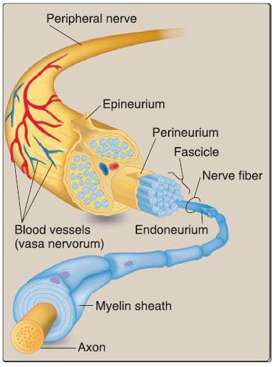
Figure 8 :Peripheral nerve structure
1. Cells: Neuroglia of the PNS include the Schwann cell and the satellite cell. A Schwann cell may produce a myelin sheath around an axon, thereby forming a myelinated axon. The junction between adjacent myelinating Schwann cells is called the node of Ranvier (see Fig. 7). In addition, a Schwann cells may produce cytoplasmic extensions around an axon, thereby forming an unmyelinated axon. The satellite cell surrounds the neuronal perikarya located within sensory, sympathetic, and parasympathetic ganglia.
2. Connective tissue components: The connective tissue components of a spinal nerve hold the axons within a spinal nerve together in bundles (see Fig. 8). The epineurium is a dense irregular connective tissue that surrounds the entire peripheral nerve (e.g., the sciatic nerve). The perineurium is a specialized type of connective tissue that surrounds a bundle of axons called a fascicle. The endoneurium is a delicate, loose connective tissue that surrounds an individual axon.
E. Autonomic nervous system
The ANS has visceromotor and viscerosensory components (although traditionally only the visceromotor component has been emphasized). The hypothalamus has central control of the ANS, coordinating all ANS actions. The ANS can be subdivided into sympathetic (thoracolumbar) and parasympathetic (craniosacral) divisions.
1. Sympathetic (thoracolumbar; T1-L2'L3) division: The visceromotor component of the sympathetic nervous system has a fight or flight or catabolic function that is necessary in emergency situations in which the body needs a sudden burst of energy, and the whole system tends to "go off together" (Fig. 9). It is a two-neuron chain that consists of a preganglionic sympathetic neuron and a postganglionic sympathetic neuron following this general pattern: short preganglionic neuron ➔ ganglion ➔ long postganglionic neuron ➔ effector organ. The exception to this arrangement is found in the sympathetic splanchnic nerves , where the pattern is long preganglionic neuron ➔ prevertebral ganglion ➔ short postganglionic neuron ➔ effector organ. The viscerosensory component carries visceral pain sensation from nociceptors located in viscera to the CNS (Fig. 10). Nociceptors are free nerve endings that respond to pathologic stimuli such as myocardial infarction, appendicitis, and gastrointestinal cramping or bloating. Visceral pain sensation is carried almost exclusively by the viscerosensory component of the sympathetic nervous system. It is poorly localized because nociceptor density is low, nociceptor fields are large, and its projection to higher CNS levels is widespread.

Figure 9: Visceromotor component of the sympathetic system. Preganglionic sympathetic neurons (solid line; green), postganglionic sympathetic neurons (dashed line; orange). C = celiac ganglion, GSp = greater thoracic splanchnic nerve, I = inferior mesenteric ganglion, IHy = inferior hypogastric plexus, LSp = lesser thoracic splanchnic nerve, LTSp = least thoracic splanchnic nerve, PARA = paravertebral chain ganglia, PRE = prevertebral ganglia, S = superior mesenteric ganglion,SCG = superior cervical ganglion, Shy= superior hypogastric plexus, Sp = splanchnic nerve.
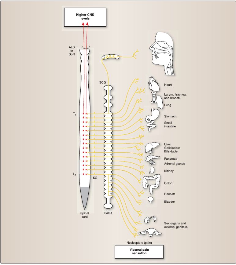
Figure 10 :Viscerosensory component of the sympathetic system (visceral pain sensation). The sensory neuron (yellow) has its perikaryon in the spinal ganglia (SG). This neuron sends a peripheral process to the viscera that ends as a free nerve ending (or nociceptor) and sends a central process into the spinal cord, which synapses with a neuron within the spinal cord (red). This neuron (red) projects axons to higher CNS levels. ALS = anterolateral system, CNS = central nervous system, PARA = paravertebral chain ganglia, SCG = superior cervical ganglion, SpR = spinoreticular tract.
2. Parasympathetic (craniosacral; CNs Ill, VII, IX, and X and SrS4)division: The visceromotor component of the parasympathetic nervous system has a rest and digest or anabolic function that is necessary to conserve energy, restore body resources, and get rid of wastes (Fig. 11). The whole visceromotor component of the parasympathetic nervous system does not "go off together" but, instead, specific activities are initiated when appropriate. It is a twoneuron chain that consists of a preganglionic parasympathetic neuron and a postganglionic parasympathetic neuron following this general pattern: long preganglionic neuron ➔ ganglion ➔ short postganglionic neuron ➔ effector organ. The viscerosensory component carries 1) arterial oxygen tension (P 30:J and arterial pH information from chemoreceptors, 2) blood pressure information from baroreceptors, 3) visceral pressure and movement sensation from rapidly adapting mechanoreceptors, 4) visceral stretch sensation from slowly adapting mechanoreceptors, 5) osmolarity information from osmoreceptors, and 6) temperature from internal thermal receptors (Fig. 12).
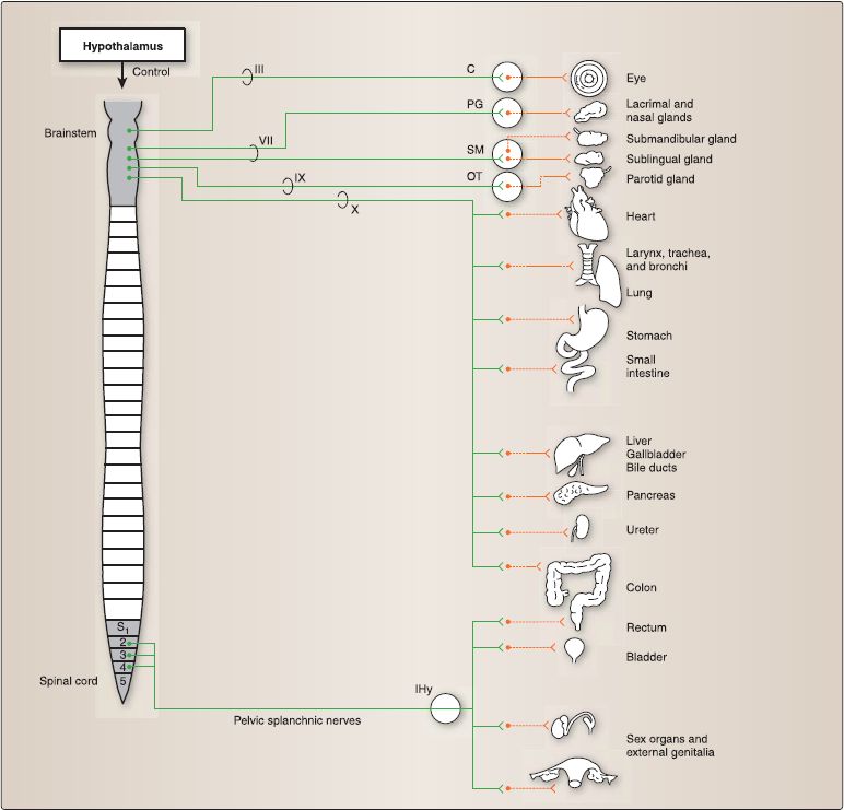
Figure 11 :Visceromotor component of the parasympathetic system. Preganglionic parasympathetic neurons (solid line; green),postganglionic parasympathetic neurons (dashed line; orange). C = ciliary ganglion, PG = pterygopalatine ganglion,SM = submandibular ganglion, OT = otic ganglion, IHy = inferior hypogastric plexus, cranial nerves Ill, VII, IX, X.

Figure 12 : Viscerosensory component of the parasympathetic system. The sensory neuron (yellow) has its perikaryon in the posterior root ganglion, geniculate ganglion (GG) of CN VII, inferior (petrosal) ganglion (IG) of CN IX, or the inferior (nodose) ganglion {IG) of CN X. This neuron sends a peripheral process to the viscera that ends as various receptors and sends a central process into the spinal cord or solitary nucleus (SN), which synapses with a neuron within the spinal cord or the SN (red).These neurons (red) project axons to higher CNS levels. CN = cranial nerve, CNS = central nervous system, SG = spinal ganglia.
Radiologic anatomy utilizes diagnostic medical imaging to study the structure and function of the human body. Diagnostic images are included throughout the text to illustrate normal and pathologic anatomy in a clinical context. Combining gross anatomical dissections, schematic illustrations, and diagnostic medical images promotes integration of gross and clinical anatomy topics. Early exposure to radiologic anatomy not only aids in the understanding of gross anatomical relationships, but also improves the learner's understanding of foundational radiology concepts and terms and familiarizes the learner with basic radiologic modalities.
A. Modalities
The following types of imaging modalities are presented throughout the text.
1. X-ray/plain film: Images are formed by x-rays that are passed through tissue onto a film/detector. This modality allows for excellent penetration of all structures and good imaging of bone and lungs. Soft tissues like fat and muscle are not well visualized on plain film.
2. Ultrasound (US): Sound waves are passed through tissue to produce an image based on the rate of reflection back to the transducer. This modality allows for excellent distinction between cystic and solid structures, but does not penetrate through bone or gas.
3. Computed tomography (CT): Three-dimensional images are rendered from a data set produced by a rotating x-ray beam. This modality allows for excellent visualization of soft tissue, gas, bone, and large vessels.
4. Magnetic resonance imaging (MRI): Three-dimensional images are produced by radio waves passed through tissue in an extremely powerful magnetic field. This modality allows for excellent imaging of bones and soft tissues (CNS, musculoskeletal system).
B. Orientation
Once the modality has been identified, develop a systematic approach to looking at radiographs. First, orientation to the image is key, and information about radiographic position, projection, and view is provided to aid in this task. Keeping these basic terms and concepts in mind will help the learner establish correct image orientation and, in turn, better appreciate the anatomy pictured in the radiologic images within each chapter.
1. Position: Position refers to the general position of the patient or the side of the patient closest to the film/detector. For this text, assume the patient is upright, unless otherwise noted (e.g., recumbent, supine, prone).
2. Projection: Projection refers to the direction of x-rays in relation to the patient and film/detector (Fig. 1.36). Common projection terms include the following:
a. Anteroposterior (A/P) projection: The x-rays travel from an anterior position, through the patient to reach the x-ray film/ detector that is posterior to the patient.
b. Posteroanterior (P/A) projection: The x-rays travel from a posterior position, through the patient to reach the film/detector that is anterior to the patient.
c. Lateral projection: The x-ray travels from one side, through the patient to reach the film/detector on the contralateral side (e.g., left to right).
3. View: The most common descriptor noted in the text, view provides information about plane and axis orientation.
a. Anteroposterior view: An A/P view describes the image as if the viewer is facing the patient; thus, the left side of the image coincides with the right side of the viewer's body.
b. Lateral view: A lateral view describes the image as if the viewer is looking at the image from the side, rather than from the front.
c. Views in CT and MRI: For CT and MRls, the specific plane is commonly included to aid in orientation to structures. For example, an MRI of the knee joint is typically viewed from a sagittal view to best visualize the internal structures (e.g., cruciate ligaments). CT images are commonly described as axial to denote an image through the long axis of the body (in the transverse plane), although CT images can also be rendered in the sagittal and coronal planes. When viewing an axial CT image, remember that it is an inferior view, meaning that the patient's feet are coming out of the plane of the image and the head into the plane of the image. Therefore, the left side of the image corresponds to the right side of the patient.
 الاكثر قراءة في علم التشريح
 اخر الاخبار
اخبار العتبة العباسية المقدسة

الآخبار الصحية


 قسم الشؤون الفكرية يصدر كتاباً يوثق تاريخ السدانة في العتبة العباسية المقدسة
 "المهمة".. إصدار قصصي يوثّق القصص الفائزة في مسابقة فتوى الدفاع المقدسة للقصة القصيرة
 (نوافذ).. إصدار أدبي يوثق القصص الفائزة في مسابقة الإمام العسكري (عليه السلام)


