آخر المواضيع المضافة

 النبات
الحيوان
الأحياء المجهرية
علم الأمراض
التقانة الإحيائية
التقنية الحيوية المكروبية
التقنية الحياتية النانوية
علم الأجنة
الأحياء الجزيئي
علم وظائف الأعضاء
الغدد
المضادات الحيوية
النبات
الحيوان
الأحياء المجهرية
علم الأمراض
التقانة الإحيائية
التقنية الحيوية المكروبية
التقنية الحياتية النانوية
علم الأجنة
الأحياء الجزيئي
علم وظائف الأعضاء
الغدد
المضادات الحيوية| Translation Occurs by Initiation,Elongation, and Termination |


|
|
Read More
Date: 24-5-2021
Date: 29-11-2015
Date: 18-5-2021
|
Translation Occurs by Initiation,Elongation, and Termination
KEY CONCEPTS
- The ribosome has three tRNA-binding sites.
- An aminoacyl-tRNA enters the A site.
- Peptidyl-tRNA is bound in the P site.
- Deacylated tRNA exits via the E site.
- An amino acid is added to the polypeptide chain by transferring the polypeptide from peptidyl-tRNA in the P site to aminoacyl-tRNA in the A site.
An amino acid is brought to the ribosome by an aminoacyl-tRNA. Its addition to the growing polypeptide chain occurs by an interaction with the tRNA that brought the previous amino acid. Each of these tRNAs lies in its own distinct site on the ribosome. Figure1 shows that the two sites have different features:
- Except for the initiator tRNA, an incoming aminoacyl-tRNA binds to the A site. Prior to the entry of aminoacyl-tRNA, the site exposes the mRNA codon representing the next amino acid to be added to the chain.
- The codon representing the most recent amino acid to have been added to the nascent polypeptide chain lies in the P site. This site is occupied by peptidyl-tRNA, a tRNA carrying the nascent polypeptide chain.
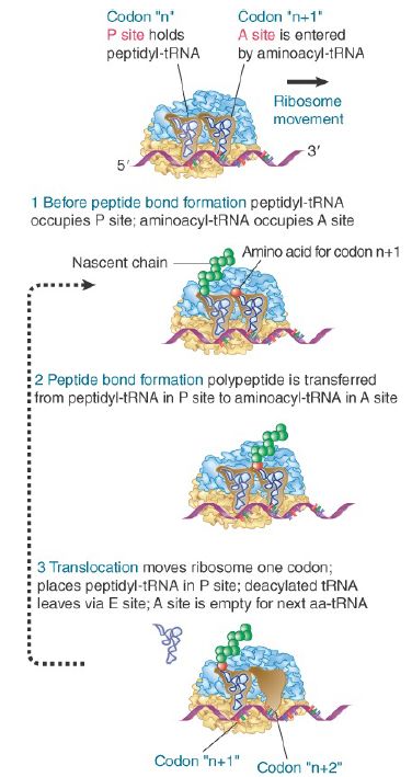
FIGURE 1. The ribosome has two sites for binding charged tRNA.
Figure 2 shows that the aminoacyl end of the tRNA is located on the large subunit, whereas the anticodon at the other end of the tRNA interacts with the mRNA bound by the small subunit. Thus, the P and A sites each extend across both ribosomal subunits.
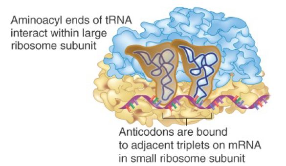
FIGURE 2. The P and A sites position the two bound tRNAs across both ribosomal subunits.
For a ribosome to form a peptide bond, it must be in the state shown in step 1 in Figure 1, when peptidyl-tRNA is in the P site and aminoacyl-tRNA is in the A site. Peptide bond formation occurs when the polypeptide carried by the peptidyl-tRNA is transferred to the amino acid carried by the aminoacyl-tRNA. This step requires correct positioning of the aminoacyl-ends of the two tRNAs within the large subunit. This reaction is catalyzed by the large subunit of the ribosome.
Transfer of the polypeptide generates the ribosome shown in step 2 of Figure 1, in which the deacylated tRNA, lacking any amino acids, lies in the P site, and a new peptidyl-tRNA is in the A site. The peptide on this peptidyl-tRNA is one amino acid residue longer than the one that was carried on the peptidyl-tRNA that had been in the P site in step 1.
The ribosome now moves one triplet along the messenger RNA. This stage is called translocation. The movement transfers the deacylated tRNA out of the P site and moves the peptidyl-tRNA into the P site (see step 3 in Figure 1). The next codon to be translated now lies in the A site, ready for a new aminoacyl-tRNA to enter, when the cycle will be repeated. Figure 3 summarizes the interaction between tRNAs and the ribosome.
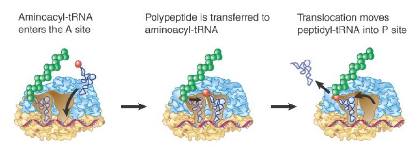
FIGURE 3. Aminoacyl-tRNA enters the A site, receives the polypeptide chain from peptidyl-tRNA, and is transferred into the P site for the next cycle of elongation.
The deacylated tRNA leaves the ribosome via another tRNA-binding site, the E site. This site is transiently occupied by the tRNA en route between leaving the P site and being released from the ribosome into the cytosol. Thus, the route of tRNA through the ribosome is into the A site, through the P site, and out through the E site . Figure 4 compares the movement of tRNA and mRNA, which may be considered a sort of ratchet in which the reaction is driven by the codon–anticodon interaction.
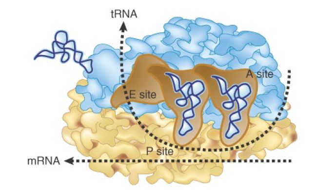
FIGURE 4. tRNA and mRNA move through the ribosome in the same direction.
Translation is divided into the three stages shown in Figure 5:
- Initiation involves the reactions that precede formation of the peptide bond between the first two amino acids of the polypeptide. It requires the ribosome to bind to the mRNA, which forms an initiation complex that contains the first aminoacyl-tRNA. This is a relatively slow step in translation and usually determines the rate at which an mRNA is translated.
- Elongation includes all the reactions from the formation of the first peptide bond to the addition of the last amino acid. Amino acids are added to the chain one at a time; the addition of an amino acid is the most rapid step in translation.
- Termination encompasses the steps that are needed to release the completed polypeptide chain; at the same time, the ribosome dissociates from the mRNA.
Different sets of accessory protein factors assist the ribosome at each stage. Energy is provided at various stages by the hydrolysis of guanine triphosphate (GTP).
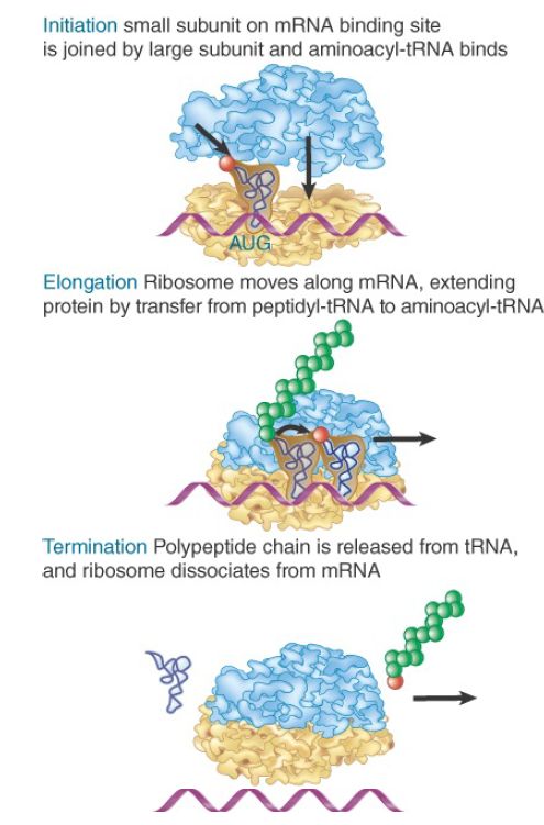
FIGURE 5 Translation has three stages.
During initiation, the small ribosomal subunit binds to mRNA and then is joined by the large subunit. During elongation, the mRNA moves through the ribosome and is translated in nucleotide triplets. (Although the ribosome is usually referred to as moving along mRNA, it is more accurate to say that the mRNA is pulled through the ribosome.) At termination, the polypeptide is released, the mRNA is released, and the individual ribosomal subunits dissociate
and can be used again.


|
|
|
|
دراسة: إجراء واحد لتقليل المخاطر الجينية للوفاة المبكرة
|
|
|
|
|
|
|
"الملح والماء" يمهدان الطريق لأجهزة كمبيوتر تحاكي الدماغ البشري
|
|
|
|
|
|
بالصور: عند زيارته لمعهد نور الإمام الحسين (عليه السلام) للمكفوفين وضعاف البصر في كربلاء.. ممثل المرجعية العليا يقف على الخدمات المقدمة للطلبة والطالبات
|
|
|
|
ممثل المرجعية العليا يؤكد استعداد العتبة الحسينية لتبني إكمال الدراسة الجامعية لشريحة المكفوفين في العراق
|
|
|
|
ممثل المرجعية العليا يؤكد على ضرورة مواكبة التطورات العالمية واستقطاب الكفاءات العراقية لتقديم أفضل الخدمات للمواطنين
|
|
|
|
العتبة الحسينية تستملك قطعة أرض في العاصمة بغداد لإنشاء مستشفى لعلاج الأورام السرطانية ومركز تخصصي للتوحد
|