Skeletal System
The skeletal system is divided into the axial skeleton and the appendicular skeleton (Fig. 1). The axial skeleton consists of bones of the cranium (or skull), hyoid bone, ribs, sternum, vertebrae, and sacrum. The appendicular skeleton consists of the bones of the upper and lower limbs, shoulder girdle, and pelvic girdle. The skeleton is composed of cartilage and bone.
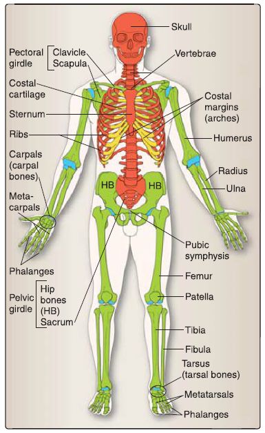
Figure 1. : Skeletal system comprises the axial skeleton (red) and the appendicular
skeleton (green) as well as articular (blue) and costal (yellow) cartilage.
A. Cartilage
The three types of cartilage include hyaline cartilage, elastic cartilage, and fibrocartilage.
1. Hyaline cartilage: Hyaline cartilage is found in fetal skeletal tissue, epiphyseal growth plates, articular surface of synovial joints, costal cartilages, nasal cartilage, laryngeal cartilage, tracheal cartilage rings, and bronchial cartilage plates. Its main features are cells, including chondrogenic cells, chondroblasts, and chondrocytes; a ground substance, containing proteoglycans (e.g., aggrecan, decorin, biglycan, and fibromodulin), hyaluronan, multiadhesive glycoproteins (e.g., chondronectin, tenascin), and water (interstitial fluid); and fibers, including types 11, VI, IX, X, and XI collagen (Fig. 2).
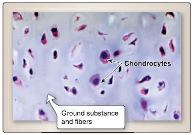
Figure 2: Hyaline cartilage.
2. Elastic cartilage: Elastic cartilage is found in the pinna of the external ear, external auditory meatus, auditory tube, epiglottis, corniculate cartilage of the larynx, and cuneiform cartilage of the larynx. Its main features are similar to those of hyaline cartilage (Fig. 3). However, the distinguishing feature of elastic cartilage is the presence of elastic fibers along with type II collagen fibers. Elastic cartilage is generally stained with a special stain (i.e., Verhoeff) that colors elastic fibers black.
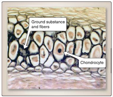
Figure 3: Elastic cartilage.
3. Fibrocartilage: Fibrocartilage is found in intervertebral disks, symphysis pubis, articular disks of the temporomandibular and sternoclavicular joints, menisci of the knee joint, and insertion of tendons. Its main features are similar to that of hyaline cartilage (Fig. 4). However, the distinguishing features of fibrocartilage include the absence of a perichondrium, more extracellular matrix than cells, and the presence of types I and II collagen fibers.
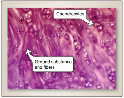
Figure 4 : Fibrocartilage.
B. Bone
Bones can be classified according to their shape. Long bones are tubular (e.g., humerus), and short bones are cuboidal (e.g., bones of the wrist and ankle). Flat bones serve a protective function (e.g., bones of the cranium). Irregular bones have various shapes (e.g., bones of the face), and sesamoid bones form in certain tendons (e.g., patella bone of the knee). Visual inspection of a bone reveals various bone markings, created by the attachment of tendons, ligaments, and fascia or by the close proximity of an artery to the bone or where an artery enters the bone, and bone formations caused by the passage of a tendon to a joint to improve its leverage. The main features of bone are cells (osteoprogenitor cells, osteoblasts, osteocytes, and osteoclasts), a ground substance (proteoglycans, hyaluronan, multiadhesive glycoproteins, vitamin K-dependent proteins, and a mineral component of hydroxyapatite crystals [Ca10 (PO4)6 OH2]), and fibers that include types I and V collagen.
1. Lamellar bone: Comprising virtually all bone in a healthy adult, lamellar bone is therefore sometimes referred to as mature bone. (Woven bone comprises bone during embryonic development, bone remodeling, and bone repair and is therefore sometimes referred to as immature bone.) Lamellar bone (compared with woven bone) is characterized by an ordered arrangement of osteocytes, a reduced amount of ground substance, a regular parallel arrangement of collagen fibers organized into lamellae or layers, and increased mineralization. It is divided into two types: compact bone and spongy bone (Fig. 5).
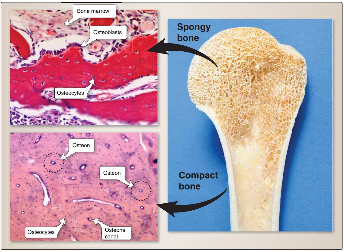 Figure 5: Spongy and compact bone.
a. Compact bone: As shown in Figure 1.9, compact bone consists predominately of osteons (or Haversian systems). The osteon is the basic unit of compact bone and consists of an osteonal (Haversian) canal and perforating (Volkmann) canals through which blood vessels and nerves travel, concentric lamellae (layers) of mineralized extracellular matrix that surround the osteonal canal, collagen fibers that are oriented in a parallel arrangement within a single lamella, osteocytes whose cell bodies reside in lacunae and whose cell processes extend through canaliculi, and a cement line that surrounds each osteon.
b. Spongy bone: Spongy bone consists predominately of a meshwork of internal struts called trabeculae (see Fig. 5). The lamellae in spongy bone are arranged in a stacked pattern (i.e., one layer on top of another layer), rather than in the concentric pattern found in the osteon of compact bone.


