Integumentary System
The integumentary system consists of the skin and epidermal derivatives (or epidermal appendages). Skin forms the outer covering of the body and is the largest organ of the body, accounting for 15%-20% of the total body weight. Epidermal derivatives include sweat and sebaceous glands, hair and hair follicles, and nails.
A. Functions
Collectively, skin and epidermal derivatives regulate body temperature and water loss, provide a nonspecific barrier to external environmental factors (e.g., microorganisms), synthesize vitamin D, absorb ultraviolet (UV) irradiation, convey sensory information, play a role in antigen presentation, and secrete sweat and sebum.
B. Skin
In general, skin consists of three layers, outer epidermis, middle dermis, and deep hypodermis or subcutaneous layer.
1. Outer epidermis: The outer epidermis is an epithelial layer classified as a keratinized stratified squamous epithelium. On the basis of the comparative thickness of the epidermis, skin is classified as either thick skin (found on palms of the hands or soles of the feet) or thin skin (covering the rest of the body), as shown in Figures 1. and 2.

Figure 1. : Thick skin. AD = adipose tissue,DER = dermis, DP = dermal papillae, EPI = epidermis, ER = epidermal ridge, ESD = eccrine sweat gland duct, ESG = eccrine sweat gland, HYP = hypodermis, PC = Pacinian corpuscle.
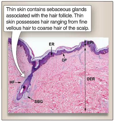
Figure 2: Thin skin. DER = dermis,DP = dermal papillae, EPI = epidermis,ER = epidermal ridge, HF = hair follicle,SBG = sebaceous gland.
2. Dermis: The junction between the epidermis and dermis is irregular, whereby epidermal ridges protrude into the underlying dermis, and dermal papillae protrude into the overlying epidermis (see Fig. 1.). The dermis is composed of the papillary layer and reticular layer. The superficial papillary layer (i.e., the dermal papillae) consists of loose connective tissue with fibroblasts, types I and Ill collagen fibers, and thin elastic fibers. The deeper reticular layer consists of dense, irregular connective tissue with fibroblasts, type I collagen, and thick elastic fibers. In addition, skin contains a number of epidermal derivatives (also called epidermal appendages): eccrine and apocrine sweat glands, sebaceous glands, hair follicles (and arrector pili muscles), and nails.
C. Epidermal derivatives
1. Eccrine sweat glands and duct: These simple, coiled tubular glands are involved in the secretion of water, electrolytes, urea, and ammonium. The duct opens onto the skin surface as sweat pores. The eccrine sweat glands function in the regulation of body temperature and emotional sweating.
2. Apocrine sweat glands and duct: These simple, coiled tubular glands are involved in the secretion of proteins, carbohydrates, ammonia, lipid, and organic compounds. The duct opens onto the skin surface in the axilla, mons pubis, and anal regions. The apocrine sweat glands function in the production of a malodorous body scent.
3. Sebaceous glands and duct: These simple acinar glands are involved in the secretion of sebum (i.e., lipid and cell debris). The short duct opens into the upper portion of a hair follicle into the pilosebaceous canal. The sebaceous glands function in the lubrication of the skin and play a role in teenage acne.
4. Hair follicles: These form as epidermal cells and grow into the underlying dermis during early embryonic development. The deepest part of the hair follicle becomes round-shaped and is called the hair bulb. The hair bulbs are invaginated by connective tissue called the dermal papillae, which are infiltrated by blood vessels and nerve endings. Epidermal cells within the hair bulb form an area containing epidermal stem cells called the germinal matrix.
The continuous proliferation and differentiation of germinal matrix cells at the tip of the dermal papilla is responsible for the formation and growth of the hair shaft, a long, slender filament that extends above the surface of the epidermis.
5. Nail: The nail is a translucent plate (called the nail plate) of closely compacted hard keratin formed by the proliferation and keratinization of epithelial cells within the nail matrix. The nail matrix is a V-shaped area located under a fold of skin called the proximal nail fold. The only portion of the nail matrix that is grossly visible is the lunula, a half moon-shaped whitish area. At the outer edge of the proximal nail fold is the eponychium or cuticle. The nail rests on top of the nail bed. At the fingertip, the nail and the nail bed fuse to form the hyponychium, which protects the nail matrix from bacterial and fungal invasion. The dermis beneath the nail bed is highly vascular, which contributes to the pink color seen through the nail, and is a clinically useful indicator of the degree of oxygenation of blood.


