آخر المواضيع المضافة

 النبات
الحيوان
الأحياء المجهرية
علم الأمراض
التقانة الإحيائية
التقنية الحيوية المكروبية
التقنية الحياتية النانوية
علم الأجنة
الأحياء الجزيئي
علم وظائف الأعضاء
الغدد
المضادات الحيوية
النبات
الحيوان
الأحياء المجهرية
علم الأمراض
التقانة الإحيائية
التقنية الحيوية المكروبية
التقنية الحياتية النانوية
علم الأجنة
الأحياء الجزيئي
علم وظائف الأعضاء
الغدد
المضادات الحيوية| Free Connective Tissue Cells-Macrophages |


|
|
Read More
Date: 18-1-2017
Date: 25-7-2016
Date: 23-1-2017
|
Free Connective Tissue Cells-Macrophages
Connective tissue macrophages are also called histiocytes, sessile macrophages or resting migratory cells. They are part of the mononuclear phagocyte system (MPS; reticuloendothelial system, RES). The precursor cells of connective tissue macrophages are monocytes, which are derive d from bone marrow. Their characteristic attribute is their ability to phagocytose and store substances (scavenger cells ). Phagocytes have irregular shapes, they are flattened and of ten show pseudopodia-like processes. Their eccentrically located nuclei are smaller and more densely structure d than fibroblast nuclei. The cytoplasmic organization is particularly striking in electron micrographs. Apart from the usual cell organelles, there are small vesicles, vacuoles, filaments and osmiophilic inclusions. These may be primary lysosomes, secondary lysosomes 1 or of ten also inclusions, which have b ecome part of phagolysosomes or residual bodies . Therefore, light microscopy of ten reveals a “granular cytoplasm” in macrophages. In-vivo injection of a vital dye (trypan blue, lithium carmine, Indian ink) will selectively stain macro-phages, because macrophages opsonize these substances and store them in their cytoplasm in the form of granules. The granules and the phagocytosed substances can be recognized in light microscopy. Macrophage from human subcutaneous connective tissue.
1 Lysosomes
2 Collagen fibrils cut vertical to their axis
Electron microscopy; magnification: × 15 000

This electron-microscopic image shows the characteristic attributes of human macrophages. During periods of very active phagocytosis, macro phages (“large scavenger cells”) show a diameter of about 20 μm and are in-deed voluminous. Their cytoplasmic processes may look like the tentacles of pseudopodia or like hook s. In the vicinity of their kidney-shape d nuclei 1 are crista-type mitochondria 2 . The granular endoplasmic reticulum (rER) is poorly developed. In contrast, note the elaborately developed Golgi apparatus 3 as well as the osmiophilic granules, which can be identified either as lysosomes or as phagosomes. Note the cross-section of a centriole . Macrophages are able to move around in the connective tissue like ameba. Therefore, they are also called wandering (mobile) phagocytes.
1 Nucleus
2 Mitochondria
3 Golgi apparatus
Electron microscopy; magnification: × 12000

Human macrophages can be isolated from subcutaneous connective tissue and grown in cell culture like the macrophage seen here. The flat, sprawling cell shows a characteristic surface relief. In the central region, it forms a dome over the cell nucleus. In this central area, the cytoplasmic processes appear relatively cloudy. However, the raised, partially branched microfolds (microplicae ) in the periphery create a veil-like edge. The microfolds can be of different lengths.
Scanning electron microscopy; magnification: × 4300

References
Kuehnel, W.(2003). Color Atlas of Cytology, Histology, and Microscopic Anatomy. 4th edition . Institute of Anatomy Universitätzu Luebeck Luebeck, Germany . Thieme Stuttgart · New York .


|
|
|
|
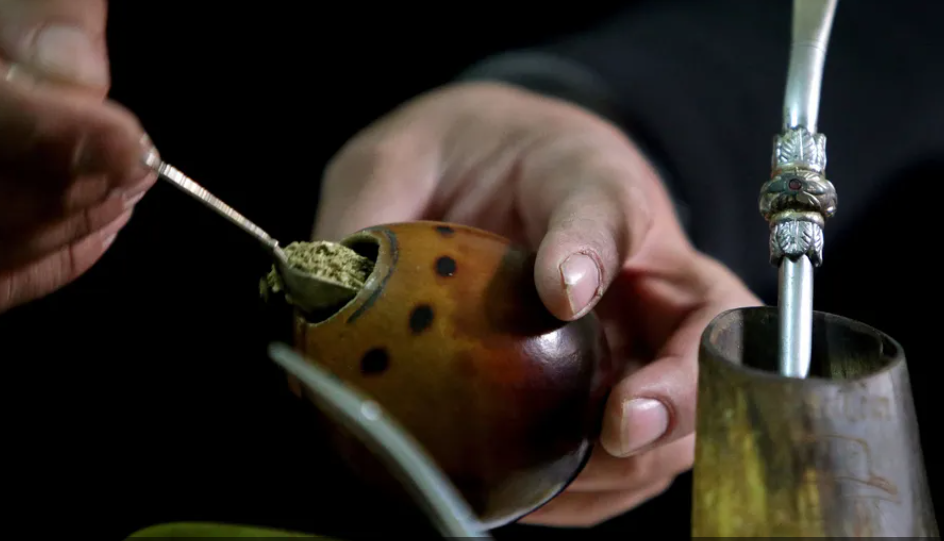
"إنقاص الوزن".. مشروب تقليدي قد يتفوق على حقن "أوزيمبيك"
|
|
|
|
|
|
|
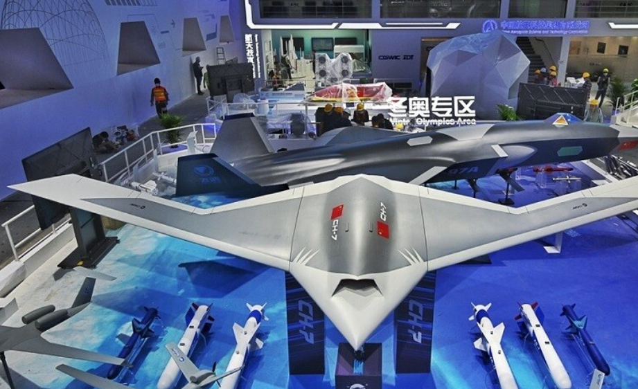
الصين تحقق اختراقا بطائرة مسيرة مزودة بالذكاء الاصطناعي
|
|
|
|
|
|
|
العتبة العباسية المقدسة تطلق النسخة الحادية عشرة من مسابقة الجود العالمية للقصيدة العمودية
|
|
|