
النبات

مواضيع عامة في علم النبات

الجذور - السيقان - الأوراق

النباتات الوعائية واللاوعائية

البذور (مغطاة البذور - عاريات البذور)

الطحالب

النباتات الطبية


الحيوان

مواضيع عامة في علم الحيوان

علم التشريح

التنوع الإحيائي

البايلوجيا الخلوية


الأحياء المجهرية

البكتيريا

الفطريات

الطفيليات

الفايروسات


علم الأمراض

الاورام

الامراض الوراثية

الامراض المناعية

الامراض المدارية

اضطرابات الدورة الدموية

مواضيع عامة في علم الامراض

الحشرات


التقانة الإحيائية

مواضيع عامة في التقانة الإحيائية


التقنية الحيوية المكروبية

التقنية الحيوية والميكروبات

الفعاليات الحيوية

وراثة الاحياء المجهرية

تصنيف الاحياء المجهرية

الاحياء المجهرية في الطبيعة

أيض الاجهاد

التقنية الحيوية والبيئة

التقنية الحيوية والطب

التقنية الحيوية والزراعة

التقنية الحيوية والصناعة

التقنية الحيوية والطاقة

البحار والطحالب الصغيرة

عزل البروتين

هندسة الجينات


التقنية الحياتية النانوية

مفاهيم التقنية الحيوية النانوية

التراكيب النانوية والمجاهر المستخدمة في رؤيتها

تصنيع وتخليق المواد النانوية

تطبيقات التقنية النانوية والحيوية النانوية

الرقائق والمتحسسات الحيوية

المصفوفات المجهرية وحاسوب الدنا

اللقاحات

البيئة والتلوث


علم الأجنة

اعضاء التكاثر وتشكل الاعراس

الاخصاب

التشطر

العصيبة وتشكل الجسيدات

تشكل اللواحق الجنينية

تكون المعيدة وظهور الطبقات الجنينية

مقدمة لعلم الاجنة


الأحياء الجزيئي

مواضيع عامة في الاحياء الجزيئي


علم وظائف الأعضاء


الغدد

مواضيع عامة في الغدد

الغدد الصم و هرموناتها

الجسم تحت السريري

الغدة النخامية

الغدة الكظرية

الغدة التناسلية

الغدة الدرقية والجار الدرقية

الغدة البنكرياسية

الغدة الصنوبرية

مواضيع عامة في علم وظائف الاعضاء

الخلية الحيوانية

الجهاز العصبي

أعضاء الحس

الجهاز العضلي

السوائل الجسمية

الجهاز الدوري والليمف

الجهاز التنفسي

الجهاز الهضمي

الجهاز البولي


المضادات الميكروبية

مواضيع عامة في المضادات الميكروبية

مضادات البكتيريا

مضادات الفطريات

مضادات الطفيليات

مضادات الفايروسات

علم الخلية

الوراثة

الأحياء العامة

المناعة

التحليلات المرضية

الكيمياء الحيوية

مواضيع متنوعة أخرى

الانزيمات
Small Saphenous Vein
المؤلف:
Kuehnel, W
المصدر:
Color Atlas of Cytology, Histology, and Microscopic Anatomy
الجزء والصفحة:
17-1-2017
1977
Small Saphenous Vein
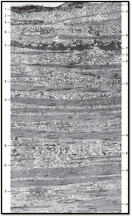
Vertical section through the wall of the small saphenous vein (vena saphena parva) at the knee joint in a 42-year-old man. The characteristic three-layered structure, as seen in the muscular arteries, is barely visible in veins. The layers of connective tissue and smooth muscle cells in venous walls are woven together without noticeable, distinct borders. In addition, the venous part of the circulatory system displays a con siderable morphological variability, which usually is an adaptation to the location of the vein in the body. The walls of the veins of the upper body, for example, consist mostly of connective tissue and only a very thin layer of muscle cells. In contrast, the musculature in the venous walls of the lower body is much more developed. This accommodates the larger hydrostatic workload for the leg and pelvic veins. Veins with few muscles occur in parts of the body where the flow regulation via lumen changes is unnecessary, e.g., in the brain, the retina and the sinuses of the dura mater. On the other hand, there are veins with strong muscular walls in organs with strong fluctuations in blood flow, such as the corpora cavernosa and the nasal mucous mem-branes. This electron micrograph shows the nucleate d sections of two flat, sprawling endothelial cells 1 . The layer immediately underneath contains the delicate fibrils of the lamina propria intimae 2 . Two spindle-shaped fibrocytes 3 are present between the collagen fibrils. These are followed by cross-sections of slender myocytes 4 and collagen fibrils in several alternating layers 5 and 6 . As always, the myocytes display their typical filament structure, even at this magnification. In the third, fibrilrich connective tissue layer (starting the count from the endothelium, top), there is a darkly stained, largely continuous elastic fiber 7 with bizarre protuberances. Sporadically, very small sections of such elastic fibers 8 occur in other fibrilrich layers as well. The outer venous wall does not have a clearly delimited tunica adventitia because the collagen fibers of the venous wall connect directly with the surrounding connective tissue. Note the collagen fibril bundles are cut across their axis in most instances, i.e., almost all collagen fibrils run longitudinally in the direction of the vein. A three-dimensional reconstruction reveals that the collagen fibrils are arranged in steep spirals.
1 Endothelial cells
2 Lamina propria intimae
3 F ibrocytes
4 Myocytes
5 Collagen fibrils, longitudinal
6 Collagen fibrils, cut across the axis
7 Elastic fiber
8 Elastic fiber material
Electron microscopy; magnification: × 6200

References
Kuehnel, W.(2003). Color Atlas of Cytology, Histology, and Microscopic Anatomy. 4th edition . Institute of Anatomy Universitätzu Luebeck Luebeck, Germany . Thieme Stuttgart · New York .
 الاكثر قراءة في علم الخلية
 اخر الاخبار
اخبار العتبة العباسية المقدسة

الآخبار الصحية


 قسم الشؤون الفكرية يصدر كتاباً يوثق تاريخ السدانة في العتبة العباسية المقدسة
 "المهمة".. إصدار قصصي يوثّق القصص الفائزة في مسابقة فتوى الدفاع المقدسة للقصة القصيرة
 (نوافذ).. إصدار أدبي يوثق القصص الفائزة في مسابقة الإمام العسكري (عليه السلام)


