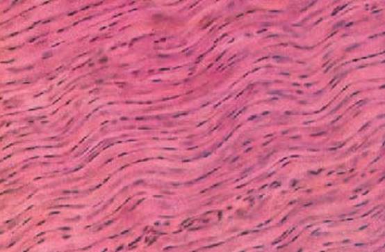
Dense Regular Connective Tissue-Tendon
The taut, dense regular connective tissue with parallel fibers is the prototype of tendons ( strand-like form ), aponeuroses ( flat, planar form) and ligaments. This figure shows part of a longitudinal section through a finger tendon. The structural components of tendons are strong, stress-absorbing collagen fibers that are commensurate with their mechanical tasks. The collagen fibers (stained red) assemble in a parallel arrangement ( primary collagen bundles). Relaxed fibers undulate slightly. Fibroblasts and their row of nuclei line up between fiber bundles. These cells are also called tendon cells ( tenocytes ). The tenocytes are in part captured in planar view and in part seen as profiles . The geometry of tendon cells adapts to the spatial conditions inside the tendon structure.
Stain: alum hematoxylin-eosin; magnification: × 240
Dense Regular Connective Tissue—Tendon
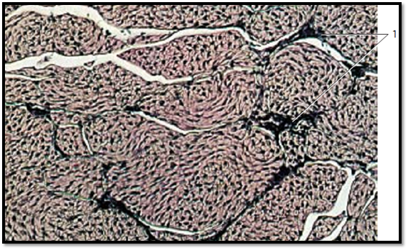
Cross-section through a tendon. Each of the fiber bundles in the tendon are encased by loose, fibrous, vascularized connective tissue ( peritendineum internum) 1 , which subdivides the tendon into secondary bundles. The tendon cells ( winged cells ) extend thin, wing-like cytoplasmic processes in all directions. The cell processes adhere closely to the collagen fibers. The light crevices are technical artifacts (shrinkage due to fixation). The tendon surface is covered by the peritendineum externum (epitendineum), which is a braided web of connective tissue. The epitendineum itself is covered at the outside with loose connective tissue called peritendineum .
1 Peritendineum internum
Stain: alum hematoxylin-eosin; magnification: × 130
Dense Regular Connective Tissue—Tendon
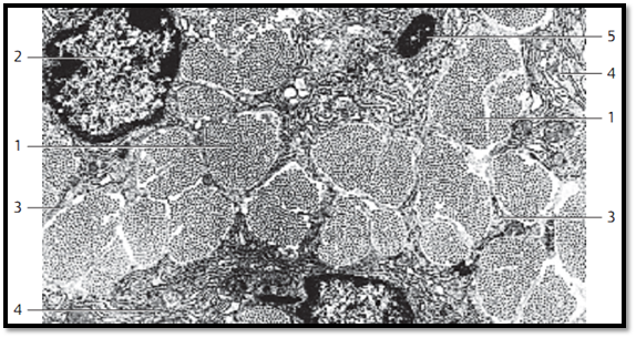
Cross-section through the tendon ( primary collagen bundle) of the long finger flexor from a mouse. Several bundles of differently sized collagen fibrils 1 are cut. In light microscopy, every bundle of fibrils corresponds to a fiber. Between the bundles of fibrils are tendon cells 2 —i.e., modified fibrocytes ( tenocytes ). Their slender cytoplasmic processes ex-tend wing-like into the narrow spaces between fibril bundles (“winged cells”) 3 . These cytoplasmic processes interconnect with each other. Tendon cells contain plenty of granular endoplasmic reticulum (rER) 4 , mitochondria and lysosomes 5 .
1 Fibril bundles
2 Nuclei of tendon cells
3 Cytoplasmic processes of tendon cells
4 Cytoplasm of tendon cells with rER
5 Lysosomes
Electron microscopy; magnification: × 8300
Dense Regular Connective Tissue—Tendon
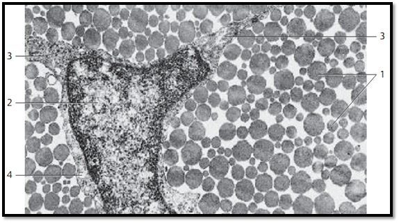
This cross-section through the tendon of the human long finger flexor out-lines the wide range for the diameters of collagen fibrils 1 . There is also a sec-tion through part of a tendon cell ( fibrocyte, tenocyte ) with its nucleus 2 and long, extended, slender processes (wings) 3 , which adhere snugly to the fibrils. Tendon cells usually have long, extended nuclei and little cytoplasm. Note the narrow perinuclear band of cytoplasm 4 .
1 Collagen fibrils, cut vertical to their axis
2 Nucleus of a tendon cell
3 Cytoplasmic processes (wing)
4 Small cytoplasmic band (seam)
Electron microscopy; magnification: × 20000
Dense Regular Connective Tissue—Tendon
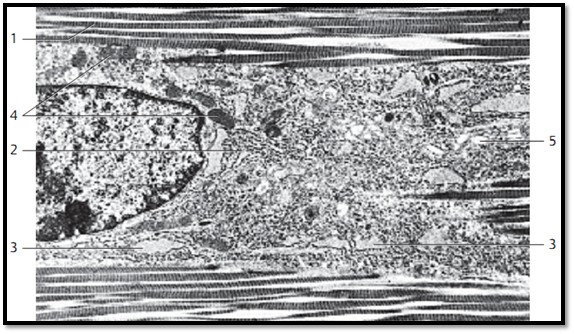
Longitudinal section through the tendon (primary collagen bundle)from the long finger flexor of a mouse. The collagen fibrils 1 are cut lengthwise and show the typical cross-striation . The section shows the perikaryon of a tendon cell ( fibrocyte, tenocyte ) between fibril bundles. The cytoplasm contains rER lamella 2 and in some sections, small rER cisternae 3 .It also contains mitochondria 4 and Golgi complexes 5 . Aponeuroses are planar tendons. Their structures resemble that of collagen ligaments.
1 Collagen fibrils
2 Granular endoplasmic reticulum, rER
3 Rough ER cisternae
4 Mitochondria
5 Golgi apparatus
Electron microscopy; magnification: × 6500
References
Kuehnel, W.(2003). Color Atlas of Cytology, Histology, and Microscopic Anatomy. 4th edition . Institute of Anatomy Universitätzu Luebeck Luebeck, Germany . Thieme Stuttgart · New York .


