آخر المواضيع المضافة

 النبات
الحيوان
الأحياء المجهرية
علم الأمراض
التقانة الإحيائية
التقنية الحيوية المكروبية
التقنية الحياتية النانوية
علم الأجنة
الأحياء الجزيئي
علم وظائف الأعضاء
الغدد
المضادات الحيوية
النبات
الحيوان
الأحياء المجهرية
علم الأمراض
التقانة الإحيائية
التقنية الحيوية المكروبية
التقنية الحياتية النانوية
علم الأجنة
الأحياء الجزيئي
علم وظائف الأعضاء
الغدد
المضادات الحيوية| Fibroblasts-Fibrocytes |


|
|
Read More
Date: 31-7-2016
Date: 1-1-2021
Date: 4-1-2017
|
Fibroblasts-Fibrocytes
Fibrocytes are local (fixed) connective tissue cells. They are branched and connect to each other via cytoplasmic processes of different sizes. Other wise, the appearances of f ibrocytes differ, dependent on the type of the connective tissue and their function. In the usual sections, they attach so tightly to the surrounding connective tissue fibers that it of ten renders their cytoplasm invisible. The name fibroblast shows that the connective tissue cell has a specific functional role. Fibroblasts play an important role in the synthesis of extracellular substances ( extracellular matrix ), as in fibrillogenesis. This figure shows strongly basophilic f ibroblasts in the connective tissue of a fetal jawbone.
Stain: hemalum-eosin; magnification: × 500

Fibroblasts from the edge fog of a cell culture ( cover-glass culture ). After seeding a cell culture, the propagating cells will spread. Their spreading in a sparse, thin layer to the underside of the cover g lass allows a microscopic examination. Fibroblasts are stretched, flattened cells. Their cytoplasmic processes may look like a membrane, or form spikes. They feature large, usually oval nuclei with prominent nucleoli and display a very delicate chromatin structure. The cytoplasm appears vitreous and is only lightly stained. Occasionally, it contains small fat droplets and vacuoles. Note the cells in mitosis 1 .
Stain: methylene blue; magnification: × 400

Fibrocytes from the connective tissue of a human amnion. Some of the oval or spindle-shaped fibrocytes have long processes, which will make contact with processes from other f ibroblasts. Whole-mount preparation. Stain: Heidenhain iron hematoxylin; magnification: × 50

Fibrocyte from the epineurium of the median nerve with arcuate, slender processes 5 of different lengths. Fibrocytes tend to have the shape of a spindle and consequently, their nuclei are elongate d and of ten lobed 1 . The electron-dense, finely granulate d cytoplasm contains many small mitochondria 2 with an electron-dense, osmiophilic matrix and close to the surface, vacuoles of various sizes 4 . The granular endoplasmic reticulum 3 and the Golgi apparatus are only poorly developed. Close to the f ibrocytes—i.e., in the extracellular matrix of the connective tissue—there are multitudes of collagen fibrils 6 , which are all cut vertical to their axis. Fibroblasts and f ibrocytes are always present in connective tissue. They are important for the formation of fibers and the synthesis of nonstructural intercellular substances ( glycosaminoglycans). The cells discharge procollagen molecules into the extracellular space, which assemble to tropocollagen and finally to microfibrils. Different stimuli convert f ibrocytes back to f ibroblasts. They will then synthesize structural and nonstructural extracellular substances, for example during wound healing.
1 Lobed nucleus
2 Mitochondria
3 Granular ER
4 Vesicles
5 Cell processes in the form of tentacles and microvilli
6 Basic substance (extracellular matrix) with collagen fibrils
Electron microscopy; magnification: × 7000

References
Kuehnel, W.(2003). Color Atlas of Cytology, Histology, and Microscopic Anatomy. 4th edition . Institute of Anatomy Universitätzu Luebeck Luebeck, Germany . Thieme Stuttgart · New York .


|
|
|
|
للعاملين في الليل.. حيلة صحية تجنبكم خطر هذا النوع من العمل
|
|
|
|
|
|
|
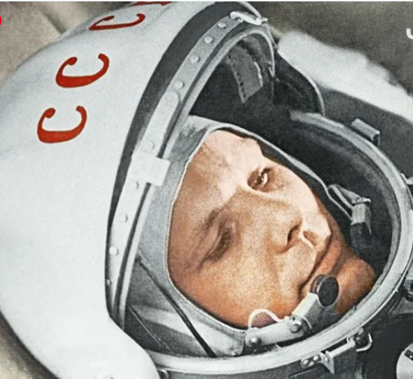
"ناسا" تحتفي برائد الفضاء السوفياتي يوري غاغارين
|
|
|
|
|
|
|
قسم الشؤون الفكرية ووفد العتبة العلوية المقدسة يبحثان سبل تعزيز التعاون في مجال الفهرسة والتوثيق
|
|
|