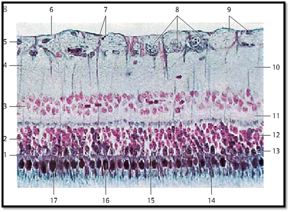
Retina
This microphotograph of a retina displays the separate layers particularly well.
1 External limiting lamina, (junctional complexes)
2 Rods and cones layer
3 Inner nuclear layer
4 Müller cells
5 Nucleus of a neuroglial cell
6 Internal limiting lamina
7 Anchor portion of a Müller cell
8 Ganglia cells of the optic nerve (3rd neuron)
9 Fiber bundle of the optic nerve
10 Inner plexiform layer (IPL)
11 Outer plexiform layer (OPL)
12 Nucleus of a rod cell
13 Nucleus of a cone cell
14 Outer segment of a rod cell
15 Inner segment of a rod cell
16 Inner segment of a cone cell
17 Outer segment of a cone cell
Stain: nuclear fast red-eosin-nigrosin (steel gray); magnification: × 320
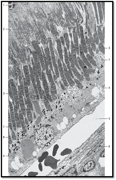
Retina
Incomplete section of a human retina. It shows the following layers:
1 External nuclear layer. This layer contains the nuclei of the rods and cones
2 Inner segments of the rods and cones with a distal acidophilic part (ellipsoid) and a proximal basophilic part (myoid). The distal ellipsoid part is filled with mitochondria, the proximal myoid part contains smooth endoplasmic reticulum membranes, Golgi complexes, and free ribosomes
3 Outer segments of the rods and cones. Their structures are in principal similar to the structure of the inner segments. Outer and inner segments of the rods are approximately of equal length. The cylindrical outer segments contain stacks of 600—1000 flat disk s, which look like rolls of coins. They are enveloped by a plasma membrane. The outer segments of the cones are cone-shaped and shorter than the outer segments of rods. They are often described as flask-shaped. The membrane stacks of cones consist of equally spaced involutions of the plasma membrane, not of separate disks. The outer third of the outer segments of the cones is surrounded by the microvilli of the pigmented epithelium 7
4 Pigment epithelium. The single-layered cuboidal epithelium is inter woven with the choroidea. Microvilli protrude from the apical portion of the epithelium. They extend far into outer segments of the rods and cones of the bacillary layer. The basal membrane borders on Bruch’s membrane and shows many folds. Pigmented epithelial cells contain large round nuclei 8 . The apical cell region contains many melanosomes and phagosomes
5 Choroidal capillary layer (lamina choroidocapillaris). This layer is found close to the pigment epithelium. It consists of a tight vascular network, which contains capillary lobes. Only one capillary is sectioned longitudinally in this figure. The capillaries contain erythrocytes
6 Lamina vasculosa. The lower right corner of the figure shows an arteriole. There are three layer s in the choroidea: the outer layer close to the sclera (Haller’s layer), the lamina vasculosa and the lamina choroidocapillaris close to the pigment epithelium.
Electron microscopy; magnification: × 3500
References
Kuehnel, W.(2003). Color Atlas of Cytology, Histology, and Microscopic Anatomy. 4th edition . Institute of Anatomy Universitätzu Luebeck Luebeck, Germany . Thieme Stuttgar t · New York .


