Starch gel electrophoresis
Historically, starch gel electrophoresis preceded agarose electrophoresis but here the order of discussion is turned about to group mechanistically related techniques. Starch gel electrophoresis was introduced by Smithies in 1955. Starch forms microreticular, thermosetting gels comprised of interlocking starch helices, cross-linked by H-bonds. The microreticular nature of starch gels introduced the phenomenon of gel sieving which revolutionized electrophoresis by greatly increasing its resolution and sensitivity.
In a microreticular gel, a protein migrating under electrophoresis faces a greatly increased frictional resistance, due to the fact that the proteins have to migrate through the 3-D gel network. This resistance is an inverse function of the size of the protein, so that small proteins will migrate with less friction than larger proteins, while proteins larger than the exclusion limit of the gel will not be able to enter into the gel at all.
Ferguson has determined that the mobility of a protein in a starch gel, as a function of the gel concentration, is described by the equation:-
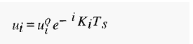
Where

i.e. the mobility decreases logarithmically as the gel concentration increases. A plot of In ui vs TS gives a straight line, of slope – t k, known as a Ferguson plot.
The same apparatus as used for paper electrophoresis and CAM-E can be used for starch gel electrophoresis. The gel is cast as a horizontal slab, which is connected to the buffer reservoirs using filter paper wicks. The slab must not be too thick to prevent excessive heat build-up. For extra cooling, the slab may be supported on a block internally cooled by circulating cold water. To accommodate the samples to be separated, small slit-like wells are cast in the gel slab, using a purpose-made mould, or cut with a scalpel. In the latter case the sample can be introduced into the slit by inserting a small strip of filter paper impregnated with sample. After sample is introduced into the sample wells, the electric field is applied across the length of the gel. Under the influence of the electric field, the sample proteins will move either towards the cathode or the anode, depending upon their charge at the buffer pH. Initially, they will migrate through the buffer in the sample wells by free electrophoresis. When they strike the well wall, on either the anodic or cathodic side. the resistance to their migration will increase and the sample will be concentrated into a narrow band.
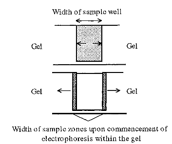
Figure 1. Sharpening of starting zones in starch gel electrophoresis.
The resolution in electrophoresis is a function of the starting bandwidth and of the band spreading during electrophoresis. The latter is largely due to diffusion of the protein from its zone of highest concentration. The structure of a microreticular gel, however, not only impedes the progress of proteins undergoing electrophoresis, brit also limits diffusion. The combination of narrow starting bands and reduced diffusion results in the marked improvements in resolution and sensitivity of gel electrophoresis. The sensitivity is increased since proteins are more easily detected the higher their concentration and, by the initial concentration of the bands and subsequent minimization of diffusion, proteins present at low levels can be detected.
A down-side of SGE is that, due to the variability of starch which is a natural product, results tend to vary from lab to lab and in the same lab at different times. This motivated a search for a more uniform, synthetic gel. Nevertheless, starch gel electrophoresis is still widely used today, mainly by biologists exploring the taxonomic relationships of organisms or in plant breeding. An advantage of starch gels is that they are non-toxic and biodegradable and are thus suitable for large-scale screening.
References
Dennison, C. (2002). A guide to protein isolation . School of Molecular mid Cellular Biosciences, University of Natal . Kluwer Academic Publishers new york, Boston, Dordrecht, London, Moscow .


