


الاخبار


اخبار الساحة الاسلامية

أخبار العتبة العلوية المقدسة
أخبار العتبة الحسينية المقدسة
أخبار العتبة الكاظمية المقدسة
أخبار العتبة العسكرية المقدسة
أخبار العتبة العباسية المقدسة
أخبار العلوم و التكنولوجيا
الاخبار الصحية
الاخبار الاقتصادية

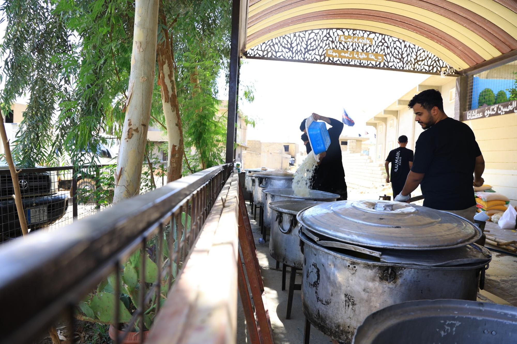
قسم الشؤون الدينية يوزع أكثر من ألفي وجبة طعام للمعزين في ذكرى عاشوراء بنينوى
 المصدر:
alkafeel.net
المصدر:
alkafeel.net
 12:13 صباحاً
 2025-07-06
 98
98


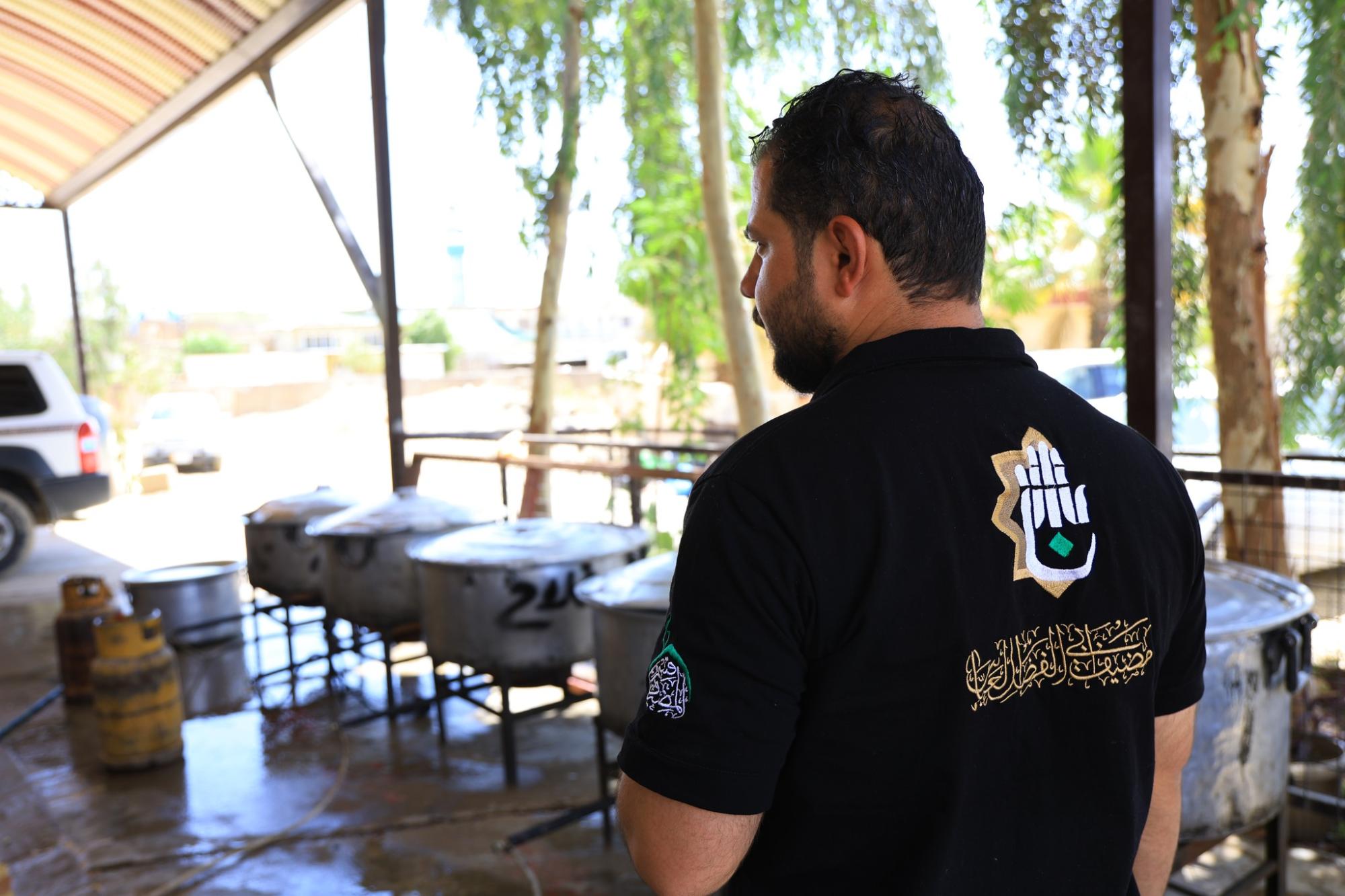
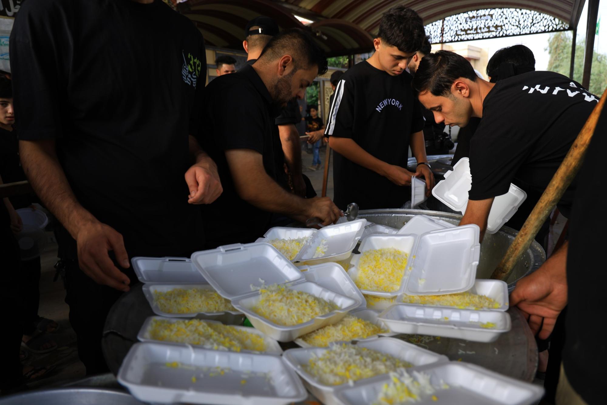
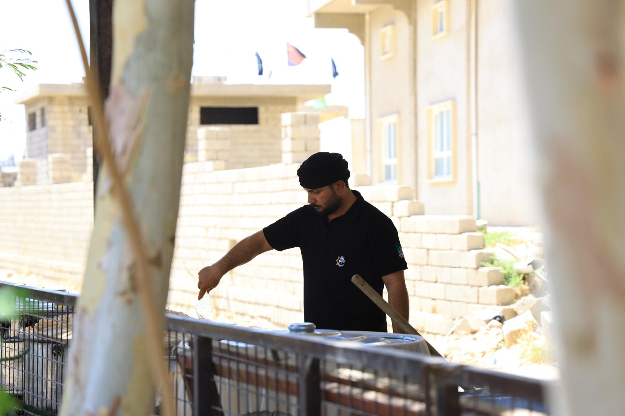
قدَّم قسم الشؤون الدينية في العتبة العباسية المقدسة أكثر من ألفي وجبة طعام للمعزين المشاركين في إحياء شهر المحرم الحرام في محافظة نينوى. وقال مسؤول شعبة الإرشاد والدعم التابعة للقسم الشيخ حيدر العارضي، إنَّ "إقامة الشعائر الحسينية في مدينة الموصل يأتي استمرارًا لنهج العتبة العباسية المقدسة في إحياء ذكرى شهادة الإمام الحسين وأهل بيته (عليهم السلام) وأصحابه". وأضاف أنَّ "الشعبة وبالتعاون مع قسم المضيف التابع للعتبة المقدسة وزعت أكثر من ألفي وجبة طعام للمعزِّين، وقد عملنا على تجهيز وجبات الطعام وتوزيعها بصورة تليق بحرمة المناسبة وأهمية الخدمة". وتابع أنَّ "إحياء عاشوراء لا يقتصر على إقامة المجالس فحسب، بل هو ميدان للبذل والعطاء في سبيل إحياء القضية الحسينية".
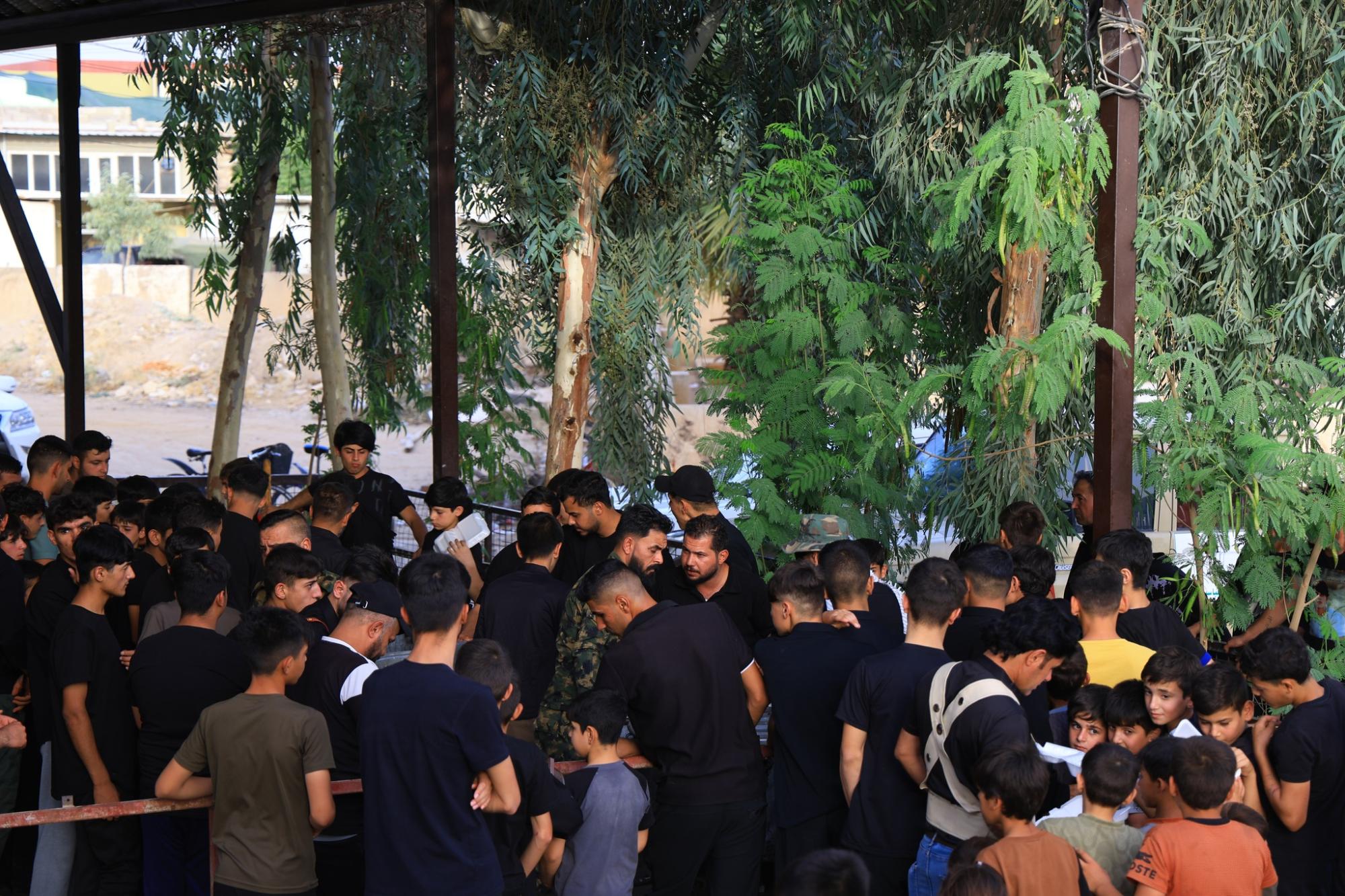
وشهدت الفعالية حضورًا واسعًا من أهالي المدينة، الذين أعربوا عن شكرهم للعتبة العباسية المقدسة، مشيدين بحسن التنظيم لمختلف الفعاليات العزائية. وتواصل شعبة الإرشاد والدعم أعمالها التبليغية والدينية في محافظة نينوى، عبر إقامة المجالس العزائية، وبالتنسيق مع المؤسسات المحلية، ضمن خطة متكاملة لإحياء شهر المحرم الحرام.
اخبار ذات صلة
 (نوافذ).. إصدار أدبي يوثق القصص الفائزة في مسابقة الإمام العسكري (عليه السلام)
 قسم الشؤون الفكرية يصدر مجموعة قصصية بعنوان (قلوب بلا مأوى)
 قسم الشؤون الفكرية يصدر مجموعة قصصية بعنوان (قلوب بلا مأوى)


