


الاخبار


اخبار الساحة الاسلامية

أخبار العتبة العلوية المقدسة
أخبار العتبة الحسينية المقدسة
أخبار العتبة الكاظمية المقدسة
أخبار العتبة العسكرية المقدسة
أخبار العتبة العباسية المقدسة
أخبار العلوم و التكنولوجيا
الاخبار الصحية
الاخبار الاقتصادية

الامين العام للعتبة الحسينية: نعمل بروح الفريق لخدمة الزائرين وتأمين مراسيم عاشوراء أولوية مطلقة
 المصدر:
imamhussain.org
المصدر:
imamhussain.org
 05:25 مساءً
 2025-07-05
 33
33


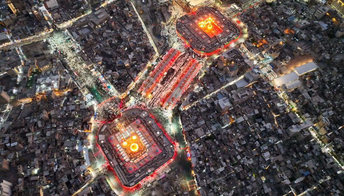 في إطار استعداداتها السنوية لاستقبال الزيارة المليونية في العاشر من محرم الحرام، أشار الأمين العام للعتبة الحسينية المقدسة الاستاذ حسن رشيد العبايجي، الى توفير أفضل الخدمات الأمنية والصحية والخدمية للزائرين القادمين من مختلف أنحاء العالم.
وقال الأمين العام للعتبة الحسينية المقدسة، "نحن كأمانة عامة للعتبة الحسينية المقدسة، هدفنا الأسمى هو تقديم أفضل الخدمات للزائرين وكذلك للهيئات والمواكب الحسينية، ودائما نطلب أن يطلق علينا لقب (خادم)، بالأمس كان هناك لقاء مع أحد الإخوة من مواكب الأطراف، فقلت له اشهد علي، ودع الإخوة يشهدون علي، أنني خادم لكم، وسجلوني خادما لكم"، مبينا أن "هدفنا هو إحياء هذه الشعائر بما يرضي سيد الشهداء (عليه السلام) وصاحب القضية، والمصيبة، لذلك نبذل أقصى الجهود خلال عاشوراء، ونسعى جاهدين في تهيئة كل متطلبات الخدمة، وراحة وسلامة الزائر، سواء من الناحية الصحية أو الأمنية أو حتى من ناحية النقل وغيرها".
وأضاف، أنه "في بداية ليلة محرم، وخلال التحضيرات لانتقال المنصة الخاصة باستقبال الزائرين وتحيتهم، دار حديث ونقاش كبير داخل أروقة إدارة العتبة الحسينية المقدسة، وبرعاية ممثل المرجعية الدينية العليا الشيخ عبد المهدي الكربلائي، وقد أخذ هذا الموضوع وقتا من النقاش، والكشف الموقعي، والزيارات الميدانية، إلى أن تم اتخاذ القرار بأن تكون المنصة خارج الصحن الشريف، وكان لهذا القرار آثار كبيرة على الجانب التنظيمي وكنا نتابع كل خطوة صغيرة ام كبيرة من اجل انجاح هذه المراسيم والاداء الجديد، فالتغيير هذا العام كان كبيرا، خلال تغيير الراية من الحمراء إلى السوداء، تم إضافة وحذف العديد من الإجراءات بهدف تقليل ازدحام الزائرين، وتهيئة مراسيم الزيارة والفرائض العبادية للزائر".
وزاد، "لقد تمكنا من استثمار كل الأفكار الممكنة، والوصول الى إخراج المنصة في الحائر الحسيني، كما تمكنا من تقليل نسبة الازدحام داخل الصحن الحسيني الشريف بنسبة تقارب (30%)، مما ساعد في تقليل التدافع الذي قد يتسبب في بعض المشاكل، كما اتخذنا إجراءات إضافية، مثل توفير السراديب للنساء لتمكينهن من أداء صلواتهن ومراسمهن بكل راحة".
وتابع، أنه "أما بالنسبة لمشاريع التوسعة التي تم تنفيذها خارج العتبة الحسينية المقدسة، فقد كانت خطوة مهمة لاستيعاب أعداد كبيرة من الزائرين، فكانت مراسيم تبديل الراية، التي تم نقلها من داخل الصحن الحسيني إلى خارجه، مما سهل على الزوار متابعة الحدث بشكل أكثر انسيابية".
وبين، أنه "من بين المشاريع التي أحدثت فارقا كبيرا، كان صحن العقيلة زينب (عليها السلام)، فلقد كانت إضافة هائلة، حيث استطاع أن يستوعب أعدادا ضخمة من الزائرين، وكنا نشعر بأن الزائرين قد استبشروا خيرا بذلك، حيث شعروا بالراحة والحرية في أداء شعائرهم دون التكدس والازدحام، وخروج المنصة واداء المراسيم (مراسيم تبديل الراية) خارج الصحن كان له اثار ايجابية كبيرة وشهد لها معظم الزوار واثنوا عليها".
وأشار إلى، أنه "فيما يتعلق بعزاء (ركضة طويريج) في يوم العاشر من محرم الحرام، فإن هذا العزاء العفوي ليس له جانب تنظيمي رسمي أو مسؤولية مباشرة من قبل جهة معينة، فالأمر ببساطة، هو تجمع الناس في منطقة (قنطرة السلام)، حيث يتوجهون إلى الإمام الحسين (عليه السلام) في أعداد هائلة قد تصل إلى (5 أو 6 مليون) زائر والسر وراء تنظيم هذا العزاء بسلاسة على الرغم من هذه الأعداد الكبيرة هو الخبرة المتراكمة على مدار الـ(20) عاما الماضية، حيث تولدت لدى إدارة العتبة الحسينية المقدسة، العديد من الحلول والمعالجات التي ساهمت في تحقيق الانسيابية في سير العزاء بشكل منتظم، بالتعاون والتنسيق مع الحكومة المحلية، وقيادة العمليات، وقيادة الشرطة، والجهات الأمنية الأخرى، فضلا عن الحشد الشعبي وفرق الطبابة".
وأوضح، أنه "بفضل هذه الخبرة والتراكمات، تم تطوير العديد من الأفكار التي ساعدت في تقليص الحلقات الزائدة أو العوائق التي قد تؤدي إلى حدوث اختناقات، فمثلا، على مستوى العتبة الحسينية المقدسة، وكما تعلمون، فإن معظم الزائرين يركزون على الدخول إلى الصحن الحسيني الشريف، مما يتطلب توفير ترتيبات إضافية لتسهيل حركة الزائرين، فمراسيم عزاء (ركضة طويريج) تتجمع فيها أعداد هائلة من الزائرين من معظم المحافظات العراقية، بالإضافة إلى القادمين من خارج العراق أيضا، لذلك، نحن على استعداد تام من الناحية الفنية، والهندسية، والأمنية، والصحية والخدمية لتوفير بيئة آمنة ومريحة للزائرين".
وأستطرد قائلا، إنه "سأتحدث عن جانب مهم وهو الأبواب، حيث لدينا أبواب دخول وأبواب خروج، أما أبواب الدخول، فهي تتطلب دقة عالية ومعايير هندسية مضبوطة لضمان راحة الزائر وسلامته، على سبيل المثال، لا يكون المنحدر شديدا، ويتم دراسة الأحمال بدقة من قبل الفريق الهندسي، بإشراف الاستاذ عبد الواحد البير، للوصول إلى مواصفات عالية، كما يتم فرش التراب وتسليط الأرض بحيث تكون مناسبة للسير، ومن المهم أن نأخذ بعين الاعتبار أن الزائرين يصلون مرهقين من الجو الحار ومعرضين لأشعة الشمس، مما يؤدي إلى تعرضهم للتعب والإرهاق، وأغلب المعزين متوسط أعمارهم بين (30 - 40) عاما أو أكثر، لذا من الضروري أن يشعر الزائر بالراحة ولا يتعرض للسقوط أو لأي عارض أثناء الدخول، حتى القلم قد يتسبب في تعثر أحدهم بسبب التعب، ولذلك أخذنا كل الاعتبارات عند تصميم أبواب الدخول".
وأشار إلى، أنه "هدفنا هو توفير انسيابية عالية في دخول الزائرين، وكذلك خروجهم بكل راحة وأمان، كما أن النزول عادة يكون أسرع من الصعود، ولذلك قمنا بتوزيع الأبواب على النحو التالي، أبواب الدخول (باب الرجاء، وباب القبلة، وباب الزينبية)، أبواب الخروج، (باب قاضي الحاجات، وباب الشهداء، وباب الكرامة، وباب السلام)".
ولفت إلى، أنه "من هذه الأبواب، يتوجه الزائر إلى منطقة ما بين الحرمين الشريفين، ولكن هناك بعض العقبات التي نحرص على تجاوزها لحماية الزائر، لأن سلامة الزائر هي أولويتنا القصوى، نحن نبذل أقصى الجهود ونسعى دائما لتقديم أفضل الأفكار والابتكارات من أجل ضمان سلامة الزائر، فهدفنا هو أن يصل الزائر إلى صحن الإمام الحسين (عليه السلام) بسهولة ويسر، دون أي معوقات أو مخاطر".
وأكد، أن "ممرات الدخول إلى الأبواب، مثل باب الرجاء، لا نسمح بأن تكون بعرض يزيد عن (4.7) متر ويبدأ المدخل من شارع الجمهورية وصولا إلى باب الرجاء، ويتم تصميم الممرات بمستوى وقياسات دقيقة لضمان انسيابية الدخول، أما باب القبلة، فقد تم تحديد العرض بمقياس (7.5) متر، حيث لا نسمح بدخول المواكب أو الزائرين بأكثر من هذا القياس مثل (8 أو 9) متر، لأنه قد يؤدي إلى حدوث اختناقات ومشاكل في انسيابية الحركة".
في الجانب الأمني قال الأمين العام للعتبة الحسينية المقدسة، إن "هناك نحو (2000) كاميرا منتشرة في مختلف الأماكن، تستخدم فيها تقنيات الذكاء الصناعي للكشف عن الوجوه المشبوهة أو تلك التي تحمل أهدافا خبيثة أو سيئة، وتتيح هذه الكاميرات باستخدام الذكاء الاصطناعي، والكشف بسرعة ودقة عن أي تهديد محتمل".
وأضاف، أنه "في الجانب الخدمي، والهندسي والفني، تم تظليل مسافة تبلغ (15) الف متر مربع، باستخدام قماش (الساران)، حيث يشمل التظليل أنواعا ثابتة ومتحركة، بالإضافة إلى ذلك، قامت شعبة التبريد التابعة لقسم المشاريع الهندسية والفنية في العتبة الحسينية المقدسة، بتركيب مظلات ضخمة تغطي مساحة تصل إلى (135) الف متر مربع، وذلك لتوفير الظل للزائرين في المساحات الواقعة خارج الحرم الحسيني الشريف، كما تم تركيب مرشات ومدافع الرذاذ لتوفير أجواء رطبة وتخفيف درجات الحرارة، فيما الحرارة والرطوبة داخل الصحن الحسيني الشريف قد تم حسابها بعناية وفقا لدراسات هندسية دقيقة".
وزاد، "كما قامت الشعبة بتوفير أجهزة ومعدات بكميات كبيرة غطت مساحات واسعة، في صحن العقيلة زينب (عليها السلام)، وصحن الإمام الحسن المجتبى (عليه السلام)، ومنطقة بين الحرمين الشريفين، وصحن الرسول الأعظم (صلى الله عليه وآله وسلم)، حيث أن مراوح الرذاذ غطت مساحة (160) الف متر مربع، فضلا هن تركيب منظومات الضباب التي غطت مساحة (145) الف متر مربع، مما ساهم في تحسين الأجواء وتخفيف درجات الحرارة".
في الجانب الخدمي، أشار الأمين العام للعتبة الحسينية المقدسة، إن "معمل هبة الوارث التابع للعتبة الحسينية المقدسة يقوم بتوزيع كؤوس الماء، بالإضافة إلى توزيع الثلج بمعدل يتراوح بين (650 إلى 1450) قالبا يوميا، على المواكب والهيئات الخدمية، أما مضيف الإمام الحسين (عليه السلام)، فيقوم بتوزيع الوجبات يوميا من (3000 إلى 4000) وجبة في الصباح، ومن (6000 إلى 7000) وجبة في الغداء، ومن (8000 إلى 11000) وجبة في العشاء، وفي يوم العاشر من محرم، يتم توزيع ما بين (20.000 إلى 27.000 ) وجبة".
وفيما يتعلق بالجانب الصحي، الذي توليه العتبة الحسينية المقدسة اهتماما كبيرا، كشف الأمين العام عن "توفير أفضل الخدمات الصحية باستخدام أحدث الأجهزة، بعضها ثابت والآخر ميداني، حيث يحمل المسعفون تجهيزات كاملة ومتطورة للفحص والعلاج، وفي حال حدوث أي مضاعفات للمريض، يتم نقله مباشرة إلى المراكز الطبية الثابتة أو المستشفيات المتنقلة التي تم افتتاحها لتلبية احتياجات الزائرين، كما تتوفر العناية الصحية المركزة في الميدان من خلال وجود مفارز طبية ومراكز طوارئ، يعمل فيها حوالي (350) من الكوادر الطبية من العراقيين والأجانب، بالإضافة إلى (350) مسعفا متطوعا مختصا في إنقاذ الحياة".
اخبار ذات صلة
 (نوافذ).. إصدار أدبي يوثق القصص الفائزة في مسابقة الإمام العسكري (عليه السلام)
 قسم الشؤون الفكرية يصدر مجموعة قصصية بعنوان (قلوب بلا مأوى)
 قسم الشؤون الفكرية يصدر مجموعة قصصية بعنوان (قلوب بلا مأوى)


