


الاخبار


اخبار الساحة الاسلامية

أخبار العتبة العلوية المقدسة
أخبار العتبة الحسينية المقدسة
أخبار العتبة الكاظمية المقدسة
أخبار العتبة العسكرية المقدسة
أخبار العتبة العباسية المقدسة
أخبار العلوم و التكنولوجيا
الاخبار الصحية
الاخبار الاقتصادية

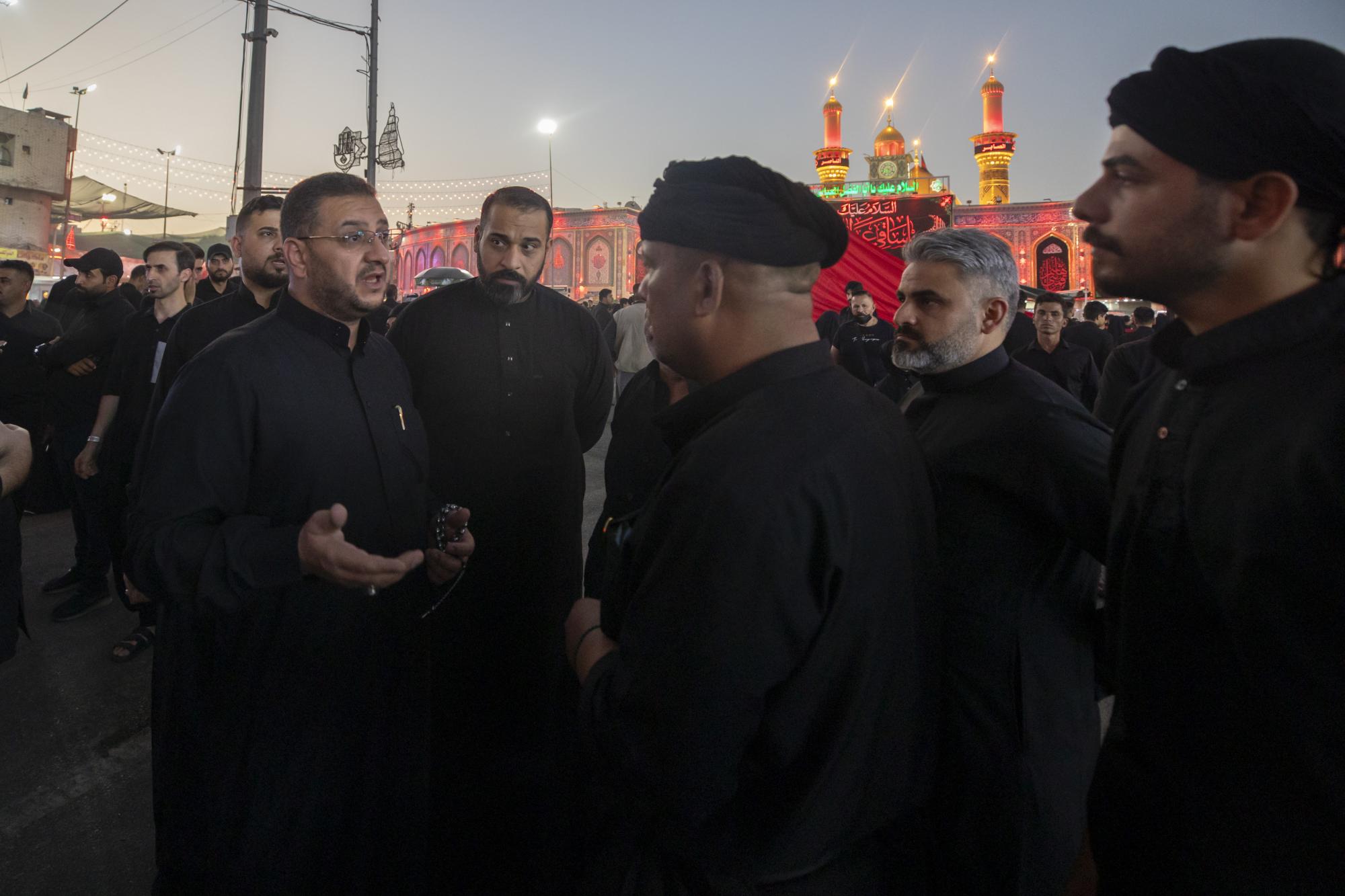
جولة تفقدية لمتابعة خطة تنظيم مداخل المعزّين المشاركين في عزاء ركضة طويريج
 المصدر:
alkafeel.net
المصدر:
alkafeel.net
 10:00 صباحاً
 2025-07-03
 93
93


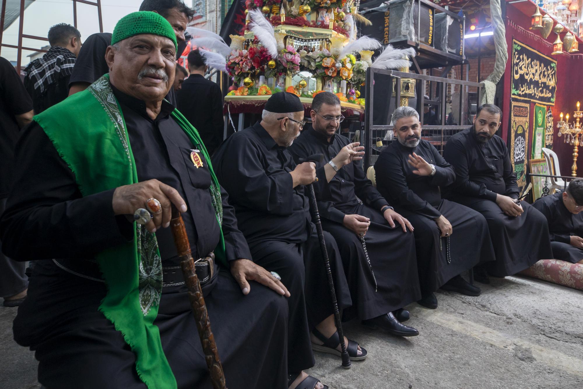
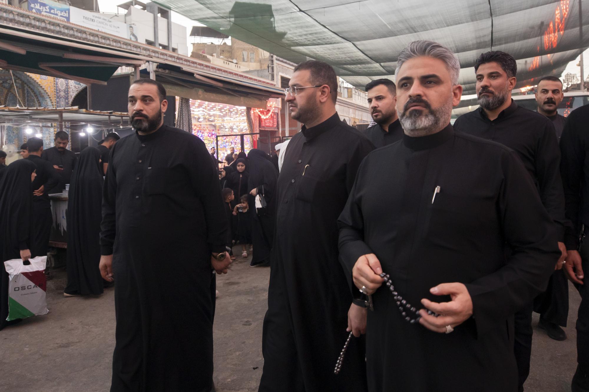
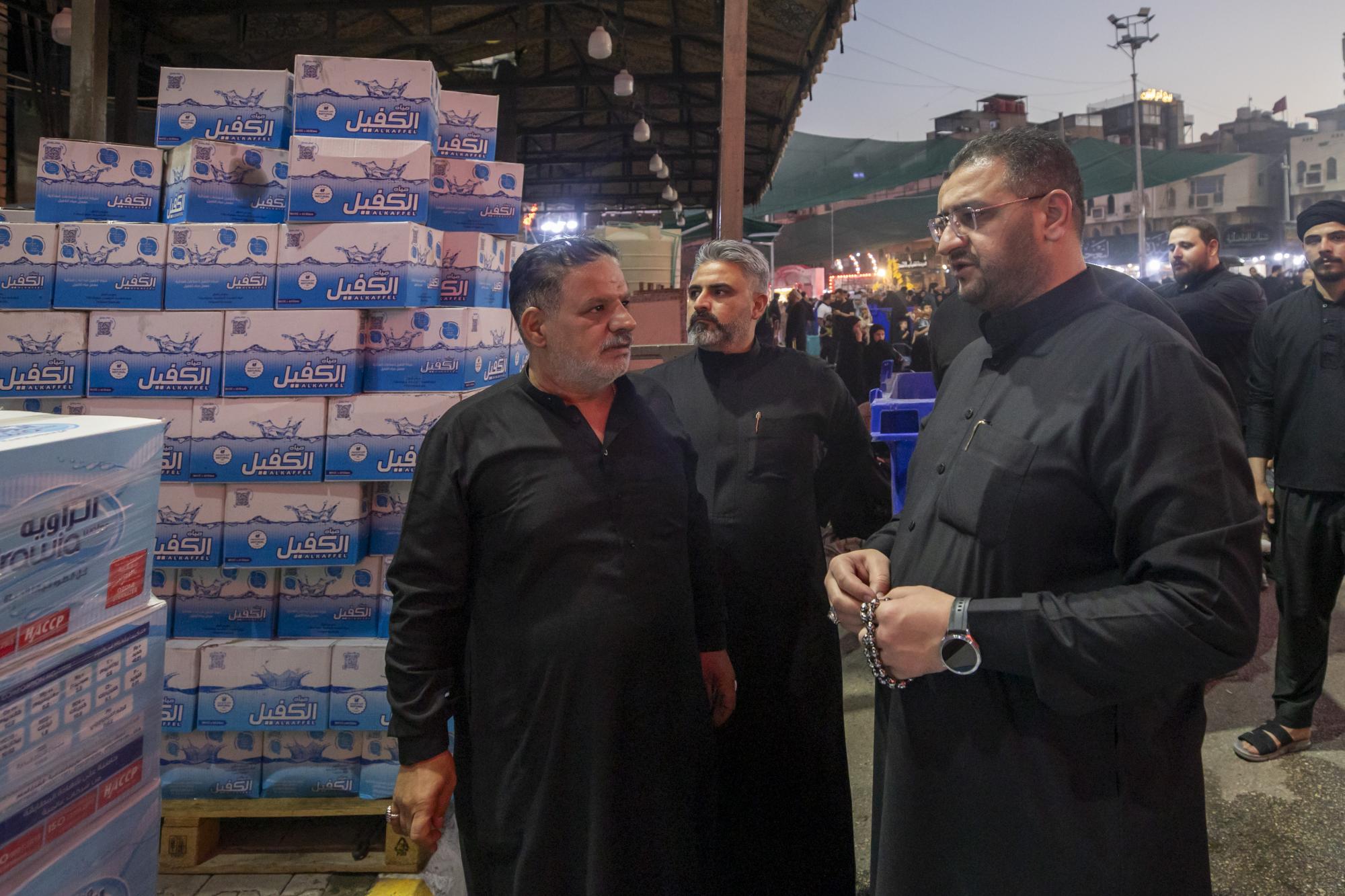
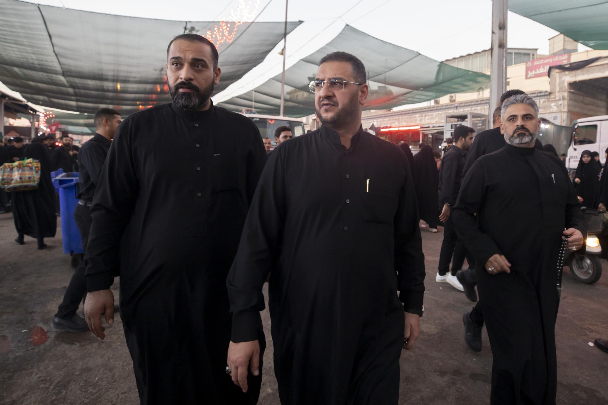
أجرى رئيس قسم حفظ النظام في العتبة العباسية المقدسة السيد حسين الشهرستاني مع رئيس قسم بين الحرمين الشريفين في العتبة المقدسة السيد حسام حسين، جولةً تفقدية لمتابعة خطة تنظيم مداخل المعزّين المشاركين في عزاء ركضة طويريج. وتهدف الجولة إلى توفير أفضل سبل الراحة والانسيابية للزائرين، فضلًا عن تسهيل حركة مرور المعزّين وتنظيم دخولهم بما يضمن سلامتهم. وتمّ في الجولة تحديد المسارات الخاصة بعزاء الركضة ووضع الترتيبات التنظيمية المناسبة، إضافةً إلى زيارة عدد من المواكب والهيآت الحسينيّة للاطّلاع على احتياجاتها والعمل على تلبيتها ضمن خطة خدمية متكاملة.
وتأتي هذه الجهود ضمن الخطة العامّة التي وضعتها العتبة العباسية المقدسة لاستقبال جموع الزائرين والمعزّين في شهر المحرم الحرام، لا سيّما في يوم العاشر الذي يشهد ذروة التوافد للمشاركة في عزاء ركضة طويريج.

اخبار ذات صلة
 (نوافذ).. إصدار أدبي يوثق القصص الفائزة في مسابقة الإمام العسكري (عليه السلام)
 قسم الشؤون الفكرية يصدر مجموعة قصصية بعنوان (قلوب بلا مأوى)
 قسم الشؤون الفكرية يصدر مجموعة قصصية بعنوان (قلوب بلا مأوى)


